Movie
Movie Controller


+ Open data

- Basic information
| Entry | Database: PDB / ID: 6ui6 | ||||||
|---|---|---|---|---|---|---|---|
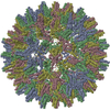
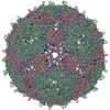
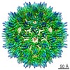
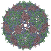
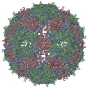
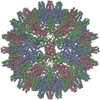
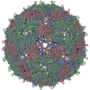
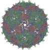
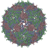
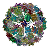
| Title | HBV T=3 149C3A | ||||||
 Components Components | Core protein | ||||||
 Keywords Keywords | VIRUS / HBV | ||||||
| Function / homology |  Function and homology information Function and homology informationmicrotubule-dependent intracellular transport of viral material towards nucleus / T=4 icosahedral viral capsid / viral penetration into host nucleus / host cell / host cell cytoplasm / symbiont entry into host cell / structural molecule activity / DNA binding / RNA binding Similarity search - Function | ||||||
| Biological species |   Hepatitis B virus Hepatitis B virus | ||||||
| Method | ELECTRON MICROSCOPY / single particle reconstruction / cryo EM / Resolution: 3.53 Å | ||||||
 Authors Authors | Wu, W. / Watts, N.R. / Cheng, N. / Huang, R. / Steven, A. / Wingfield, P.T. | ||||||
 Citation Citation | Journal: PLoS Comput Biol / Year: 2020 Title: Expression of quasi-equivalence and capsid dimorphism in the Hepadnaviridae. Authors: Weimin Wu / Norman R Watts / Naiqian Cheng / Rick Huang / Alasdair C Steven / Paul T Wingfield /  Abstract: Hepatitis B virus (HBV) is a leading cause of liver disease. The capsid is an essential component of the virion and it is therefore of interest how it assembles and disassembles. The capsid protein ...Hepatitis B virus (HBV) is a leading cause of liver disease. The capsid is an essential component of the virion and it is therefore of interest how it assembles and disassembles. The capsid protein is unusual both for its rare fold and that it polymerizes according to two different icosahedral symmetries, causing the polypeptide chain to exist in seven quasi-equivalent environments: A, B, and C in AB and CC dimers in T = 3 capsids, and A, B, C, and D in AB and CD dimers in T = 4 capsids. We have compared the two capsids by cryo-EM at 3.5 Å resolution. To ensure a valid comparison, the two capsids were prepared and imaged under identical conditions. We find that the chains have different conformations and potential energies, with the T = 3 C chain having the lowest. Three of the four quasi-equivalent dimers are asymmetric with respect to conformation and potential energy; however, the T = 3 CC dimer is symmetrical and has the lowest potential energy although its intra-dimer interface has the least free energy of formation. Of all the inter-dimer interfaces, the CB interface has the least area and free energy, in both capsids. From the calculated energies of higher-order groupings of dimers discernible in the lattices we predict early assembly intermediates, and indeed we observe such structures by negative stain EM of in vitro assembly reactions. By sequence analysis and computational alanine scanning we identify key residues and motifs involved in capsid assembly. Our results explain several previously reported observations on capsid assembly, disassembly, and dimorphism. | ||||||
| History |
|
- Structure visualization
| Movie |
Movie viewer |
|---|---|
| Structure viewer | Molecule: MolmilJmol/JSmol |
- Downloads & links
-Download
| PDBx/mmCIF format | 6ui6.cif.gz | 147 KB | Display | PDBx/mmCIF format |
|---|---|---|---|---|
| PDB format | pdb6ui6.ent.gz | 119.4 KB | Display | PDB format |
| PDBx/mmJSON format | 6ui6.json.gz | Tree view | PDBx/mmJSON format | |
| Others |  Other downloads Other downloads |
-Validation report
| Arichive directory | https://data.pdbj.org/pub/pdb/validation_reports/ui/6ui6ftp://data.pdbj.org/pub/pdb/validation_reports/ui/6ui6 | HTTPS FTP |
|---|
-Related structure data
| Related structure data | 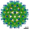 20669MC  6ui7C M: map data used to model this data C: citing same article ( |
|---|---|
| Similar structure data |


-Links
 PDBj
PDBj

- Assembly
| Deposited unit | 
|
|---|---|
| 1 | x 60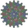
|
| 2 |
|
| 3 | x 5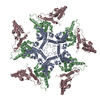
|
| 4 | x 6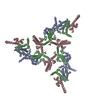
|
| 5 | 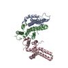
|
| Symmetry | Point symmetry: (Schoenflies symbol: I (icosahedral)) |
-Components
| #1: Protein | Mass: 16143.387 Da / Num. of mol.: 3 Source method: isolated from a genetically manipulated source Source: (gene. exp.) Hepatitis B virus / Production host:  |
|---|
-Experimental details
-Experiment
| Experiment | Method: ELECTRON MICROSCOPY |
|---|---|
| EM experiment | Aggregation state: PARTICLE / 3D reconstruction method: single particle reconstruction |
- Sample preparation
| Component | Name: Hepatitis B virus / Type: VIRUS / Entity ID: all / Source: RECOMBINANT |
|---|---|
| Source (natural) | Organism: Hepatitis B virus |
| Source (recombinant) | Organism: |
| Details of virus | Empty: YES / Enveloped: NO / Isolate: OTHER / Type: VIRION |
| Buffer solution | pH: 7.4 |
| Specimen | Embedding applied: NO / Shadowing applied: NO / Staining applied: NO / Vitrification applied: YES |
| Specimen support | Details: unspecified |
| Vitrification | Cryogen name: ETHANE |
- Electron microscopy imaging
| Experimental equipment |  Model: Tecnai Polara / Image courtesy: FEI Company |
|---|---|
| Microscopy | Model: FEI POLARA 300 |
| Electron gun | Electron source:  FIELD EMISSION GUN / Accelerating voltage: 300 kV / Illumination mode: SPOT SCAN FIELD EMISSION GUN / Accelerating voltage: 300 kV / Illumination mode: SPOT SCAN |
| Electron lens | Mode: OTHER |
| Image recording | Electron dose: 25 e/Å2 / Detector mode: COUNTING / Film or detector model: GATAN K2 SUMMIT (4k x 4k) |
- Processing
| EM software |
| ||||||||||||||||||||
|---|---|---|---|---|---|---|---|---|---|---|---|---|---|---|---|---|---|---|---|---|---|
| CTF correction | Type: PHASE FLIPPING AND AMPLITUDE CORRECTION | ||||||||||||||||||||
| 3D reconstruction | Resolution: 3.53 Å / Resolution method: FSC 0.143 CUT-OFF / Num. of particles: 15000 / Symmetry type: POINT | ||||||||||||||||||||
| Atomic model building | Protocol: FLEXIBLE FIT | ||||||||||||||||||||
| Atomic model building | PDB-ID: 1QGT Accession code: 1QGT / Source name: PDB / Type: experimental model |