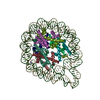
Movie
Movie Controller


+ Open data

- Basic information
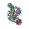
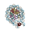
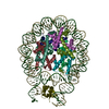
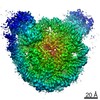
| Entry | Database: PDB / ID: 6s01 | |||||||||
|---|---|---|---|---|---|---|---|---|---|---|
| Title | Structure of LEDGF PWWP domain bound H3K36 methylated nucleosome | |||||||||
 Components Components |
| |||||||||
 Keywords Keywords | TRANSCRIPTION / LEDGF / PWWP / H3K36me3 / nucleosome | |||||||||
| Function / homology |  Function and homology information Function and homology informationIntegration of viral DNA into host genomic DNA / Autointegration results in viral DNA circles / supercoiled DNA binding / 2-LTR circle formation / Formation of WDR5-containing histone-modifying complexes / Vpr-mediated nuclear import of PICs / mRNA 5'-splice site recognition / Integration of provirus / APOBEC3G mediated resistance to HIV-1 infection / heterochromatin ...Integration of viral DNA into host genomic DNA / Autointegration results in viral DNA circles / supercoiled DNA binding / 2-LTR circle formation / Formation of WDR5-containing histone-modifying complexes / Vpr-mediated nuclear import of PICs / mRNA 5'-splice site recognition / Integration of provirus / APOBEC3G mediated resistance to HIV-1 infection / heterochromatin / nuclear periphery / euchromatin / structural constituent of chromatin / heterochromatin formation / nucleosome / nucleosome assembly / response to heat / response to oxidative stress / DNA-binding transcription factor binding / transcription coactivator activity / chromatin remodeling / protein heterodimerization activity / chromatin binding / positive regulation of transcription by RNA polymerase II / DNA binding / RNA binding / nucleoplasm / nucleus / cytosol Similarity search - Function | |||||||||
| Biological species |  Homo sapiens (human) Homo sapiens (human)synthetic construct (others) | |||||||||
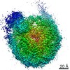
| Method | ELECTRON MICROSCOPY / single particle reconstruction / cryo EM / Resolution: 3.2 Å | |||||||||
 Authors Authors | Wang, H. / Farnung, L. / Dienemann, C. / Cramer, P. | |||||||||
| Funding support |  Germany, 2items Germany, 2items
| |||||||||
 Citation Citation | Journal: Nat Struct Mol Biol / Year: 2020 Title: Structure of H3K36-methylated nucleosome-PWWP complex reveals multivalent cross-gyre binding. Authors: Haibo Wang / Lucas Farnung / Christian Dienemann / Patrick Cramer / Abstract: Recognition of histone-modified nucleosomes by specific reader domains underlies the regulation of chromatin-associated processes. Whereas structural studies revealed how reader domains bind modified ...Recognition of histone-modified nucleosomes by specific reader domains underlies the regulation of chromatin-associated processes. Whereas structural studies revealed how reader domains bind modified histone peptides, it is unclear how reader domains interact with modified nucleosomes. Here, we report the cryo-electron microscopy structure of the PWWP reader domain of human transcriptional coactivator LEDGF in complex with an H3K36-methylated nucleosome at 3.2-Å resolution. The structure reveals multivalent binding of the reader domain to the methylated histone tail and to both gyres of nucleosomal DNA, explaining the known cooperative interactions. The observed cross-gyre binding may contribute to nucleosome integrity during transcription. The structure also explains how human PWWP domain-containing proteins are recruited to H3K36-methylated regions of the genome for transcription, histone acetylation and methylation, and for DNA methylation and repair. | |||||||||
| History |
|
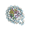
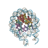
- Structure visualization
| Movie |
Movie viewer |
|---|---|
| Structure viewer | Molecule: MolmilJmol/JSmol |
- Downloads & links
-Download
| PDBx/mmCIF format | 6s01.cif.gz | 316 KB | Display | PDBx/mmCIF format |
|---|---|---|---|---|
| PDB format | pdb6s01.ent.gz | 233.6 KB | Display | PDB format |
| PDBx/mmJSON format | 6s01.json.gz | Tree view | PDBx/mmJSON format | |
| Others |  Other downloads Other downloads |
-Validation report
| Summary document | 6s01_validation.pdf.gz | 1.1 MB | Display | wwPDB validaton report |
|---|---|---|---|---|
| Full document | 6s01_full_validation.pdf.gz | 1.1 MB | Display | |
| Data in XML | 6s01_validation.xml.gz | 41.9 KB | Display | |
| Data in CIF | 6s01_validation.cif.gz | 65.1 KB | Display | |
| Arichive directory | https://data.pdbj.org/pub/pdb/validation_reports/s0/6s01ftp://data.pdbj.org/pub/pdb/validation_reports/s0/6s01 | HTTPS FTP |
-Related structure data
| Related structure data | 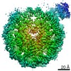 10069MC M: map data used to model this data C: citing same article ( |
|---|---|
| Similar structure data |

-Links
 PDBj
PDBj


- Assembly
| Deposited unit | 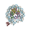
|
|---|---|
| 1 |
|
-Components
-Protein , 5 types, 9 molecules AEBFCGDHK
| #1: Protein | Mass: 15331.982 Da / Num. of mol.: 2 Source method: isolated from a genetically manipulated source Details: residue K36 was chemically modified to be ML3. / Source: (gene. exp.)  #2: Protein | Mass: 11263.231 Da / Num. of mol.: 2 Source method: isolated from a genetically manipulated source Source: (gene. exp.) #3: Protein | Mass: 13978.241 Da / Num. of mol.: 2 Source method: isolated from a genetically manipulated source Source: (gene. exp.) #4: Protein | Mass: 13524.752 Da / Num. of mol.: 2 Source method: isolated from a genetically manipulated source Source: (gene. exp.) #7: Protein | | Mass: 60224.453 Da / Num. of mol.: 1 Source method: isolated from a genetically manipulated source Details: density of residue 30-34 and residue 92-530 are invisible Source: (gene. exp.) Homo sapiens (human) / Gene: PSIP1, DFS70, LEDGF, PSIP2 / Production host:  Trichoplusia ni (cabbage looper) / References: UniProt: O75475 Trichoplusia ni (cabbage looper) / References: UniProt: O75475 |
|---|
-Wisdom 601 DNA (165- ... , 2 types, 2 molecules IJ
| #5: DNA chain | Mass: 50699.316 Da / Num. of mol.: 1 / Source method: obtained synthetically / Source: (synth.) synthetic construct (others) |
|---|---|
| #6: DNA chain | Mass: 51170.602 Da / Num. of mol.: 1 / Source method: obtained synthetically / Source: (synth.) synthetic construct (others) |
-Details
| Has ligand of interest | Y |
|---|---|
| Has protein modification | Y |
-Experimental details
-Experiment
| Experiment | Method: ELECTRON MICROSCOPY |
|---|---|
| EM experiment | Aggregation state: PARTICLE / 3D reconstruction method: single particle reconstruction |
- Sample preparation
| Component |
| |||||||||||||||||||||||||||||||||||
|---|---|---|---|---|---|---|---|---|---|---|---|---|---|---|---|---|---|---|---|---|---|---|---|---|---|---|---|---|---|---|---|---|---|---|---|---|
| Molecular weight | Value: 0.25 MDa / Experimental value: NO | |||||||||||||||||||||||||||||||||||
| Source (natural) |
| |||||||||||||||||||||||||||||||||||
| Source (recombinant) |
| |||||||||||||||||||||||||||||||||||
| Buffer solution | pH: 7.5 / Details: Solution were made from stock solution | |||||||||||||||||||||||||||||||||||
| Buffer component |
| |||||||||||||||||||||||||||||||||||
| Specimen | Conc.: 0.2 mg/ml / Embedding applied: NO / Shadowing applied: NO / Staining applied: NO / Vitrification applied: YES / Details: the complex is purified by gel filtration | |||||||||||||||||||||||||||||||||||
| Specimen support | Grid material: GOLD / Grid mesh size: 200 divisions/in. / Grid type: Quantifoil, UltrAuFoil | |||||||||||||||||||||||||||||||||||
| Vitrification | Instrument: FEI VITROBOT MARK IV / Cryogen name: ETHANE / Humidity: 100 % / Chamber temperature: 277 K / Details: blot for 4 seconds before plunging |
- Electron microscopy imaging
| Experimental equipment |  Model: Titan Krios / Image courtesy: FEI Company |
|---|---|
| Microscopy | Model: FEI TITAN KRIOS |
| Electron gun | Electron source:  FIELD EMISSION GUN / Accelerating voltage: 300 kV / Illumination mode: FLOOD BEAM FIELD EMISSION GUN / Accelerating voltage: 300 kV / Illumination mode: FLOOD BEAM |
| Electron lens | Mode: BRIGHT FIELD / Cs: 2.7 mm / C2 aperture diameter: 70 µm / Alignment procedure: COMA FREE |
| Specimen holder | Specimen holder model: FEI TITAN KRIOS AUTOGRID HOLDER |
| Image recording | Average exposure time: 9 sec. / Electron dose: 43.18 e/Å2 / Detector mode: COUNTING / Film or detector model: GATAN K2 SUMMIT (4k x 4k) |
- Processing
| EM software |
| ||||||||||||||||||||||||||||||||
|---|---|---|---|---|---|---|---|---|---|---|---|---|---|---|---|---|---|---|---|---|---|---|---|---|---|---|---|---|---|---|---|---|---|
| CTF correction | Type: PHASE FLIPPING AND AMPLITUDE CORRECTION | ||||||||||||||||||||||||||||||||
| Particle selection | Num. of particles selected: 527640 | ||||||||||||||||||||||||||||||||
| Symmetry | Point symmetry: C1 (asymmetric) | ||||||||||||||||||||||||||||||||
| 3D reconstruction | Resolution: 3.2 Å / Resolution method: FSC 0.143 CUT-OFF / Num. of particles: 55142 / Num. of class averages: 1 / Symmetry type: POINT | ||||||||||||||||||||||||||||||||
| Atomic model building | B value: 120 / Protocol: RIGID BODY FIT / Space: REAL / Target criteria: Cross-correlation coefficient | ||||||||||||||||||||||||||||||||
| Atomic model building | PDB-ID: 3MVD Accession code: 3MVD / Source name: PDB / Type: experimental model |