Movie
Movie Controller

[English] 日本語
 Yorodumi
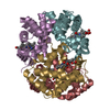
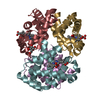
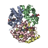
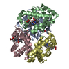
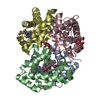
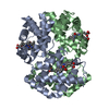
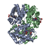
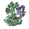
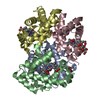
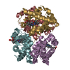
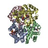
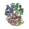
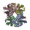
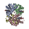
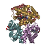
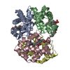
- PDB-6nbc: human methemoglobin state 1 determined using single-particle cryo... -
+ Open data

- Basic information
| Entry | Database: PDB / ID: 6nbc | ||||||
|---|---|---|---|---|---|---|---|
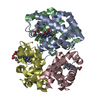
| Title | human methemoglobin state 1 determined using single-particle cryo-EM at 200 keV | ||||||
 Components Components |
| ||||||
 Keywords Keywords | OXYGEN TRANSPORT / heme-binding / hetero-4-mer / globin | ||||||
| Function / homology |  Function and homology information Function and homology informationnitric oxide transport / hemoglobin alpha binding / cellular oxidant detoxification / hemoglobin binding / haptoglobin-hemoglobin complex / renal absorption / hemoglobin complex / oxygen transport / Scavenging of heme from plasma / endocytic vesicle lumen ...nitric oxide transport / hemoglobin alpha binding / cellular oxidant detoxification / hemoglobin binding / haptoglobin-hemoglobin complex / renal absorption / hemoglobin complex / oxygen transport / Scavenging of heme from plasma / endocytic vesicle lumen / blood vessel diameter maintenance / oxygen carrier activity / hydrogen peroxide catabolic process / carbon dioxide transport / response to hydrogen peroxide / Erythrocytes take up oxygen and release carbon dioxide / Erythrocytes take up carbon dioxide and release oxygen / Heme signaling / Late endosomal microautophagy / Cytoprotection by HMOX1 / oxygen binding / platelet aggregation / regulation of blood pressure / Chaperone Mediated Autophagy / positive regulation of nitric oxide biosynthetic process / tertiary granule lumen / Factors involved in megakaryocyte development and platelet production / blood microparticle / ficolin-1-rich granule lumen / iron ion binding / inflammatory response / heme binding / Neutrophil degranulation / extracellular space / extracellular exosome / extracellular region / metal ion binding / membrane / cytosol Similarity search - Function | ||||||
| Biological species |  Homo sapiens (human) Homo sapiens (human) | ||||||
| Method | ELECTRON MICROSCOPY / single particle reconstruction / cryo EM / Resolution: 2.8 Å | ||||||
 Authors Authors | Herzik Jr., M.A. / Wu, M. / Lander, G.C. | ||||||
| Funding support |  United States, 1items United States, 1items
| ||||||
 Citation Citation | Journal: Nat Commun / Year: 2019 Title: High-resolution structure determination of sub-100 kDa complexes using conventional cryo-EM. Authors: Mark A Herzik / Mengyu Wu / Gabriel C Lander / Abstract: Determining high-resolution structures of biological macromolecules amassing less than 100 kilodaltons (kDa) has been a longstanding goal of the cryo-electron microscopy (cryo-EM) community. While ...Determining high-resolution structures of biological macromolecules amassing less than 100 kilodaltons (kDa) has been a longstanding goal of the cryo-electron microscopy (cryo-EM) community. While the Volta phase plate has enabled visualization of specimens in this size range, this instrumentation is not yet fully automated and can present technical challenges. Here, we show that conventional defocus-based cryo-EM methodologies can be used to determine high-resolution structures of specimens amassing less than 100 kDa using a transmission electron microscope operating at 200 keV coupled with a direct electron detector. Our ~2.7 Å structure of alcohol dehydrogenase (82 kDa) proves that bound ligands can be resolved with high fidelity to enable investigation of drug-target interactions. Our ~2.8 Å and ~3.2 Å structures of methemoglobin demonstrate that distinct conformational states can be identified within a dataset for proteins as small as 64 kDa. Furthermore, we provide the sub-nanometer cryo-EM structure of a sub-50 kDa protein. | ||||||
| History |
|
- Structure visualization
| Movie |
Movie viewer |
|---|---|
| Structure viewer | Molecule: MolmilJmol/JSmol |
- Downloads & links
-Download
| PDBx/mmCIF format | 6nbc.cif.gz | 888 KB | Display | PDBx/mmCIF format |
|---|---|---|---|---|
| PDB format | pdb6nbc.ent.gz | 779.7 KB | Display | PDB format |
| PDBx/mmJSON format | 6nbc.json.gz | Tree view | PDBx/mmJSON format | |
| Others |  Other downloads Other downloads |
-Validation report
| Summary document | 6nbc_validation.pdf.gz | 1.8 MB | Display | wwPDB validaton report |
|---|---|---|---|---|
| Full document | 6nbc_full_validation.pdf.gz | 1.8 MB | Display | |
| Data in XML | 6nbc_validation.xml.gz | 123.2 KB | Display | |
| Data in CIF | 6nbc_validation.cif.gz | 176.9 KB | Display | |
| Arichive directory | https://data.pdbj.org/pub/pdb/validation_reports/nb/6nbcftp://data.pdbj.org/pub/pdb/validation_reports/nb/6nbc | HTTPS FTP |
-Related structure data
| Related structure data | 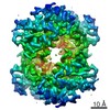 0407MC 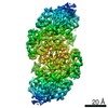 0406C 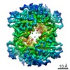 0408C 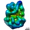 0409C 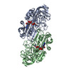 6nbbC 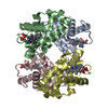 6nbdC M: map data used to model this data C: citing same article ( |
|---|---|
| Similar structure data | |
| EM raw data | EMPIAR-10250 (Title: Human methemoglobin movies obtained using Talos Arctica operating at 200 kV equipped with a K2 Data size: 1.9 TB Data #1: Raw, unaligned movie stacks of human methemoglobin acquired on a Talos Arctica using a K2 direct electron detector [micrographs - multiframe]) |
-Links
 PDBj
PDBj


- Assembly
| Deposited unit | 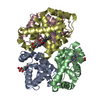
|
|---|---|
| 1 |
|
| Number of models | 4 |
-Components
| #1: Protein | Mass: 14993.159 Da / Num. of mol.: 2 / Source method: isolated from a natural source / Source: (natural) Homo sapiens (human)Plasmid details: Obtained in lyophilized form from Sigma Aldrich References: UniProt: P69905 #2: Protein | Mass: 15459.697 Da / Num. of mol.: 2 / Source method: isolated from a natural source / Source: (natural) Homo sapiens (human)Plasmid details: Obtained in lyophilized form from Sigma Aldrich References: UniProt: P68871 #3: Chemical | ChemComp-HEM /   Mass: 616.487 Da / Num. of mol.: 4 / Source method: obtained synthetically / Formula: C34H32FeN4O4 Mass: 616.487 Da / Num. of mol.: 4 / Source method: obtained synthetically / Formula: C34H32FeN4O4 |
|---|
-Experimental details
-Experiment
| Experiment | Method: ELECTRON MICROSCOPY |
|---|---|
| EM experiment | Aggregation state: PARTICLE / 3D reconstruction method: single particle reconstruction |
- Sample preparation
| Component | Name: methemoglobin from human / Type: COMPLEX Details: Lyophilized human methemoglobin purchased from Sigma Aldrich Entity ID: #1-#2 / Source: NATURAL | |||||||||||||||||||||||||
|---|---|---|---|---|---|---|---|---|---|---|---|---|---|---|---|---|---|---|---|---|---|---|---|---|---|---|
| Molecular weight | Value: 0.064 MDa / Experimental value: NO | |||||||||||||||||||||||||
| Source (natural) | Organism: Homo sapiens (human) | |||||||||||||||||||||||||
| Buffer solution | pH: 7.5 | |||||||||||||||||||||||||
| Buffer component |
| |||||||||||||||||||||||||
| Specimen | Conc.: 12 mg/ml / Embedding applied: NO / Shadowing applied: NO / Staining applied: NO / Vitrification applied: YES Details: Lyophilized human methemoglobin (Sigma Aldrich) was solubilized. | |||||||||||||||||||||||||
| Specimen support | Details: Grids were plasma cleaned using a Solarus plasma cleaner (Gatan, Inc.). Grid material: GOLD / Grid mesh size: 300 divisions/in. / Grid type: Quantifoil, UltrAuFoil, R1.2/1.3 | |||||||||||||||||||||||||
| Vitrification | Instrument: HOMEMADE PLUNGER / Cryogen name: ETHANE / Humidity: 90 % / Chamber temperature: 277.15 K Details: Sample was manually blotted for 4-5 seconds using Whatman No. 1 filter paper immediately prior to plunge-freezing. |
- Electron microscopy imaging
| Experimental equipment |  Model: Talos Arctica / Image courtesy: FEI Company |
|---|---|
| Microscopy | Model: FEI TALOS ARCTICA |
| Electron gun | Electron source:  FIELD EMISSION GUN / Accelerating voltage: 200 kV / Illumination mode: FLOOD BEAM FIELD EMISSION GUN / Accelerating voltage: 200 kV / Illumination mode: FLOOD BEAM |
| Electron lens | Mode: BRIGHT FIELD / Nominal magnification: 73000 X / Nominal defocus max: 16000 nm / Nominal defocus min: 5000 nm / Cs: 2.7 mm / C2 aperture diameter: 70 µm / Alignment procedure: COMA FREE |
| Specimen holder | Cryogen: NITROGEN / Specimen holder model: FEI TITAN KRIOS AUTOGRID HOLDER |
| Image recording | Average exposure time: 11 sec. / Electron dose: 69 e/Å2 / Detector mode: COUNTING / Film or detector model: GATAN K2 SUMMIT (4k x 4k) / Num. of grids imaged: 2 / Num. of real images: 1673 |
| Image scans | Sampling size: 5 µm / Width: 3710 / Height: 3838 / Movie frames/image: 44 / Used frames/image: 1-44 |
- Processing
| Software | Name: PHENIX / Version: 1.11.1_2580: / Classification: refinement | ||||||||||||||||||||||||||||||||||||||||
|---|---|---|---|---|---|---|---|---|---|---|---|---|---|---|---|---|---|---|---|---|---|---|---|---|---|---|---|---|---|---|---|---|---|---|---|---|---|---|---|---|---|
| EM software |
| ||||||||||||||||||||||||||||||||||||||||
| Image processing | Details: Counting mode, 250 ms frames, exposure rate of ~1.95 e- pixel-1 s-1, total exposure of ~69 e- angstrom-2 (1.57 e- angstrom-2 frame-1). | ||||||||||||||||||||||||||||||||||||||||
| CTF correction | Type: PHASE FLIPPING AND AMPLITUDE CORRECTION | ||||||||||||||||||||||||||||||||||||||||
| Particle selection | Num. of particles selected: 1232543 / Details: Difference-of-Gaussian-picked. | ||||||||||||||||||||||||||||||||||||||||
| Symmetry | Point symmetry: C2 (2 fold cyclic) | ||||||||||||||||||||||||||||||||||||||||
| 3D reconstruction | Resolution: 2.8 Å / Resolution method: FSC 0.143 CUT-OFF / Num. of particles: 24308 / Symmetry type: POINT | ||||||||||||||||||||||||||||||||||||||||
| Atomic model building | B value: 67 / Protocol: FLEXIBLE FIT / Space: REAL | ||||||||||||||||||||||||||||||||||||||||
| Atomic model building | PDB-ID: 4N7P Pdb chain-ID: A / Accession code: 4N7P / Source name: PDB / Type: experimental model | ||||||||||||||||||||||||||||||||||||||||
| Refine LS restraints |
|