 Movie
Movie Controller

[English] 日本語
 Yorodumi
- PDB-5jvv: Crystal structure and characterization an elongating GH family 16... -
+ Open data

- Basic information
| Entry | Database: PDB / ID: 5jvv | ||||||
|---|---|---|---|---|---|---|---|
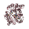
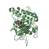
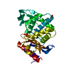
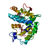
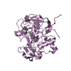
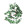
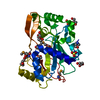
| Title | Crystal structure and characterization an elongating GH family 16 beta-1,3-glucosyltransferase | ||||||
 Components Components | beta-1,3-glucosyltransferase | ||||||
 Keywords Keywords | TRANSFERASE / glucoside hydrolase / beta-1 / 3-glucosyltransferase | ||||||
| Function / homology |  Function and homology information Function and homology informationendo-1,3(4)-beta-glucanase / cellulose catabolic process / hydrolase activity, hydrolyzing O-glycosyl compounds / side of membrane / plasma membrane Similarity search - Function | ||||||
| Biological species |  Paecilomyces sp. J18 (fungus) Paecilomyces sp. J18 (fungus) | ||||||
| Method |  X-RAY DIFFRACTION / SYNCHROTRON / MOLECULAR REPLACEMENT / Resolution: 1.589 Å X-RAY DIFFRACTION / SYNCHROTRON / MOLECULAR REPLACEMENT / Resolution: 1.589 Å | ||||||
 Authors Authors | Qin, Z. / Yan, Q. / Yang, S. / Jiang, Z. | ||||||
 Citation Citation | Journal: J. Biol. Chem. / Year: 2017 Title: Catalytic Mechanism of a Novel Glycoside Hydrolase Family 16 "Elongating" beta-Transglycosylase Authors: Qin, Z. / Yang, S. / Zhao, L. / You, X. / Yan, Q. / Jiang, Z. | ||||||
| History |
|
- Structure visualization
| Structure viewer | Molecule: MolmilJmol/JSmol |
|---|
- Downloads & links
-Download
| PDBx/mmCIF format | 5jvv.cif.gz | 260.7 KB | Display | PDBx/mmCIF format |
|---|---|---|---|---|
| PDB format | pdb5jvv.ent.gz | 209.9 KB | Display | PDB format |
| PDBx/mmJSON format | 5jvv.json.gz | Tree view | PDBx/mmJSON format | |
| Others |  Other downloads Other downloads |
-Validation report
| Summary document | 5jvv_validation.pdf.gz | 427.7 KB | Display | wwPDB validaton report |
|---|---|---|---|---|
| Full document | 5jvv_full_validation.pdf.gz | 428.7 KB | Display | |
| Data in XML | 5jvv_validation.xml.gz | 29.2 KB | Display | |
| Data in CIF | 5jvv_validation.cif.gz | 45.6 KB | Display | |
| Arichive directory | https://data.pdbj.org/pub/pdb/validation_reports/jv/5jvvftp://data.pdbj.org/pub/pdb/validation_reports/jv/5jvv | HTTPS FTP |
-Related structure data
| Related structure data | 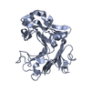 3wdtS S: Starting model for refinement |
|---|---|
| Similar structure data |


-Links
 PDBj
PDBj
- Assembly
| Deposited unit | 
| ||||||||
|---|---|---|---|---|---|---|---|---|---|
| 1 | 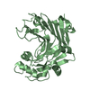
| ||||||||
| 2 | 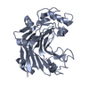
| ||||||||
| Unit cell |
|
-Components
| #1: Protein | Mass: 32540.783 Da / Num. of mol.: 2 Source method: isolated from a genetically manipulated source Source: (gene. exp.) Paecilomyces sp. J18 (fungus) / Strain: J18Production host:  Strain (production host): BL21-Gold(DE3)pLysS AG References: UniProt: A0A1S4NYE9*PLUS, Transferases; Glycosyltransferases; Hexosyltransferases #2: Water | ChemComp-HOH / |  Mass: 18.015 Da / Num. of mol.: 699 / Source method: isolated from a natural source / Formula: H2O Mass: 18.015 Da / Num. of mol.: 699 / Source method: isolated from a natural source / Formula: H2OHas protein modification | Y | |
|---|
-Experimental details
-Experiment
| Experiment | Method: X-RAY DIFFRACTION / Number of used crystals: 1 |
|---|
- Sample preparation
| Crystal | Density Matthews: 1.89 Å3/Da / Density % sol: 34.93 % |
|---|---|
| Crystal grow | Temperature: 292 K / Method: vapor diffusion, sitting drop / pH: 4.4 / Details: 35% PEG 3350, 0.1M Na-Citrate pH4.4 / PH range: 4.0-5.0 |
-Data collection
| Diffraction | Mean temperature: 100 K |
|---|---|
| Diffraction source | Source: SYNCHROTRON / Site: SSRF  / Beamline: BL19U1 / Wavelength: 0.9792 Å / Beamline: BL19U1 / Wavelength: 0.9792 Å |
| Detector | Type: DECTRIS PILATUS3 6M / Detector: PIXEL / Date: Dec 16, 2015 |
| Radiation | Protocol: SINGLE WAVELENGTH / Monochromatic (M) / Laue (L): M / Scattering type: x-ray |
| Radiation wavelength | Wavelength: 0.9792 Å / Relative weight: 1 |
| Reflection | Resolution: 1.589→37.17 Å / Num. obs: 62905 / % possible obs: 96.6 % / Redundancy: 6.9 % / Rmerge(I) obs: 0.09 / Net I/σ(I): 17.93 |
| Reflection shell | Resolution: 1.589→1.65 Å / Redundancy: 6.9 % / Rmerge(I) obs: 0.303 / Mean I/σ(I) obs: 6.11 / % possible all: 94.1 |
- Processing
| Software |
| |||||||||||||||||||||||||||||||||||||||||||||||||||||||||||||||||||||||||||||||||||||||||||||||||||||||||
|---|---|---|---|---|---|---|---|---|---|---|---|---|---|---|---|---|---|---|---|---|---|---|---|---|---|---|---|---|---|---|---|---|---|---|---|---|---|---|---|---|---|---|---|---|---|---|---|---|---|---|---|---|---|---|---|---|---|---|---|---|---|---|---|---|---|---|---|---|---|---|---|---|---|---|---|---|---|---|---|---|---|---|---|---|---|---|---|---|---|---|---|---|---|---|---|---|---|---|---|---|---|---|---|---|---|---|
| Refinement | Method to determine structure: MOLECULAR REPLACEMENT Starting model: 3WDT Resolution: 1.589→37.167 Å / SU ML: 0.12 / Cross valid method: FREE R-VALUE / σ(F): 0 / Phase error: 18.04
| |||||||||||||||||||||||||||||||||||||||||||||||||||||||||||||||||||||||||||||||||||||||||||||||||||||||||
| Solvent computation | Shrinkage radii: 0.9 Å / VDW probe radii: 1.11 Å | |||||||||||||||||||||||||||||||||||||||||||||||||||||||||||||||||||||||||||||||||||||||||||||||||||||||||
| Refinement step | Cycle: LAST / Resolution: 1.589→37.167 Å
| |||||||||||||||||||||||||||||||||||||||||||||||||||||||||||||||||||||||||||||||||||||||||||||||||||||||||
| Refine LS restraints |
| |||||||||||||||||||||||||||||||||||||||||||||||||||||||||||||||||||||||||||||||||||||||||||||||||||||||||
| LS refinement shell |
| |||||||||||||||||||||||||||||||||||||||||||||||||||||||||||||||||||||||||||||||||||||||||||||||||||||||||
| Refinement TLS params. | Method: refined / Origin x: 3.6466 Å / Origin y: -23.8877 Å / Origin z: -14.6415 Å
| |||||||||||||||||||||||||||||||||||||||||||||||||||||||||||||||||||||||||||||||||||||||||||||||||||||||||
| Refinement TLS group | Selection details: all |
