 Movie
Movie Controller


+ Open data

- Basic information
| Entry | Database: PDB / ID: 3wku | ||||||
|---|---|---|---|---|---|---|---|
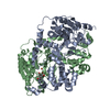
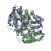
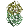
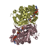
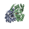
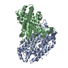
| Title | Crystal structure of the anaerobic DesB-gallate complex | ||||||
 Components Components | Gallate dioxygenase | ||||||
 Keywords Keywords | OXIDOREDUCTASE / type II extradiol dioxygenase / domain-swap dimer / extradiol dioxygenase / Fe2+ binding | ||||||
| Function / homology |  Function and homology information Function and homology informationoxidoreductase activity, acting on single donors with incorporation of molecular oxygen, incorporation of two atoms of oxygen / ferrous iron binding Similarity search - Function | ||||||
| Biological species |  Sphingobium (bacteria) Sphingobium (bacteria) | ||||||
| Method |  X-RAY DIFFRACTION / SYNCHROTRON / MOLECULAR REPLACEMENT / Resolution: 2.7 Å X-RAY DIFFRACTION / SYNCHROTRON / MOLECULAR REPLACEMENT / Resolution: 2.7 Å | ||||||
 Authors Authors | Sugimoto, K. / Senda, M. / Kasai, D. / Fukuda, M. / Masai, E. / Senda, T. | ||||||
 Citation Citation | Journal: Plos One / Year: 2014 Title: Molecular Mechanism of Strict Substrate Specificity of an Extradiol Dioxygenase, DesB, Derived from Sphingobium sp. SYK-6 Authors: Sugimoto, K. / Senda, M. / Kasai, D. / Fukuda, M. / Masai, E. / Senda, T. | ||||||
| History |
|
- Structure visualization
| Structure viewer | Molecule: MolmilJmol/JSmol |
|---|
- Downloads & links
-Download
| PDBx/mmCIF format | 3wku.cif.gz | 168.3 KB | Display | PDBx/mmCIF format |
|---|---|---|---|---|
| PDB format | pdb3wku.ent.gz | 133.7 KB | Display | PDB format |
| PDBx/mmJSON format | 3wku.json.gz | Tree view | PDBx/mmJSON format | |
| Others |  Other downloads Other downloads |
-Validation report
| Summary document | 3wku_validation.pdf.gz | 453.2 KB | Display | wwPDB validaton report |
|---|---|---|---|---|
| Full document | 3wku_full_validation.pdf.gz | 476.7 KB | Display | |
| Data in XML | 3wku_validation.xml.gz | 31.1 KB | Display | |
| Data in CIF | 3wku_validation.cif.gz | 42.1 KB | Display | |
| Arichive directory | https://data.pdbj.org/pub/pdb/validation_reports/wk/3wkuftp://data.pdbj.org/pub/pdb/validation_reports/wk/3wku | HTTPS FTP |
-Related structure data
| Related structure data | 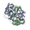 3wpmC  3wr3C  3wr4C 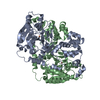 3wr8C 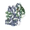 3wr9C  3wraC 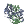 3wrbC 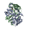 3wrcC  3vju 3vjv 3vjw 3vjx 3vjy C: citing same article ( |
|---|---|
| Similar structure data |


-Links
 PDBj
PDBj- Assembly
| Deposited unit | 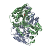
| ||||||||
|---|---|---|---|---|---|---|---|---|---|
| 1 |
| ||||||||
| Unit cell |
|
-Components
| #1: Protein | Mass: 46953.164 Da / Num. of mol.: 2 Source method: isolated from a genetically manipulated source Source: (gene. exp.) Sphingobium (bacteria) / Strain: SYK-6 / Gene: desB / Production host: #2: Chemical |   Mass: 55.845 Da / Num. of mol.: 2 / Source method: obtained synthetically / Formula: Fe Mass: 55.845 Da / Num. of mol.: 2 / Source method: obtained synthetically / Formula: Fe#3: Chemical | ChemComp-GDE / |   Mass: 170.120 Da / Num. of mol.: 1 / Source method: obtained synthetically / Formula: C7H6O5 Mass: 170.120 Da / Num. of mol.: 1 / Source method: obtained synthetically / Formula: C7H6O5#4: Water | ChemComp-HOH / |  Mass: 18.015 Da / Num. of mol.: 2 / Source method: isolated from a natural source / Formula: H2O Mass: 18.015 Da / Num. of mol.: 2 / Source method: isolated from a natural source / Formula: H2O |
|---|
-Experimental details
-Experiment
| Experiment | Method: X-RAY DIFFRACTION / Number of used crystals: 1 |
|---|
- Sample preparation
| Crystal | Density Matthews: 2.17 Å3/Da / Density % sol: 43.26 % |
|---|---|
| Crystal grow | Temperature: 277 K / Method: vapor diffusion, sitting drop / pH: 7.75 Details: 25% PEG8000, 0.1M sodium acetate, 0.1M HEPES-NaOH, pH 7.75, VAPOR DIFFUSION, SITTING DROP, temperature 277K |
-Data collection
| Diffraction | Mean temperature: 95 K |
|---|---|
| Diffraction source | Source: SYNCHROTRON / Site: Photon Factory  / Beamline: BL-5A / Wavelength: 1 Å / Beamline: BL-5A / Wavelength: 1 Å |
| Detector | Type: ADSC QUANTUM 315r / Detector: CCD / Date: Mar 12, 2010 |
| Radiation | Monochromator: Si 111 / Protocol: SINGLE WAVELENGTH / Monochromatic (M) / Laue (L): M / Scattering type: x-ray |
| Radiation wavelength | Wavelength: 1 Å / Relative weight: 1 |
| Reflection | Resolution: 2.7→56.8 Å / Num. all: 43335 / Num. obs: 42572 / % possible obs: 98.2 % / Observed criterion σ(F): 0 / Observed criterion σ(I): 0 |
| Reflection shell | Resolution: 2.7→2.85 Å / Rmerge(I) obs: 0.0309 / % possible all: 98.5 |
- Processing
| Software |
| |||||||||||||||||||||||||||||||||||||||||||||||||||||||||||||||||
|---|---|---|---|---|---|---|---|---|---|---|---|---|---|---|---|---|---|---|---|---|---|---|---|---|---|---|---|---|---|---|---|---|---|---|---|---|---|---|---|---|---|---|---|---|---|---|---|---|---|---|---|---|---|---|---|---|---|---|---|---|---|---|---|---|---|---|
| Refinement | Method to determine structure: MOLECULAR REPLACEMENT / Resolution: 2.7→20 Å / Cor.coef. Fo:Fc: 0.923 / Cor.coef. Fo:Fc free: 0.859 / SU B: 18.891 / SU ML: 0.397 / Cross valid method: THROUGHOUT / σ(F): 0 / ESU R Free: 0.481 / Stereochemistry target values: MAXIMUM LIKELIHOOD / Details: HYDROGENS HAVE BEEN ADDED IN THE RIDING POSITIONS
| |||||||||||||||||||||||||||||||||||||||||||||||||||||||||||||||||
| Solvent computation | Ion probe radii: 0.8 Å / Shrinkage radii: 0.8 Å / VDW probe radii: 1.4 Å / Solvent model: MASK | |||||||||||||||||||||||||||||||||||||||||||||||||||||||||||||||||
| Displacement parameters | Biso mean: 61.634 Å2
| |||||||||||||||||||||||||||||||||||||||||||||||||||||||||||||||||
| Refinement step | Cycle: LAST / Resolution: 2.7→20 Å
| |||||||||||||||||||||||||||||||||||||||||||||||||||||||||||||||||
| Refine LS restraints |
| |||||||||||||||||||||||||||||||||||||||||||||||||||||||||||||||||
| LS refinement shell | Resolution: 2.7→2.769 Å / Total num. of bins used: 20
|
