 Movie
Movie Controller


+ Open data

- Basic information
| Entry | Database: PDB / ID: 1yq5 | ||||||
|---|---|---|---|---|---|---|---|
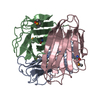
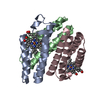
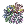
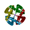
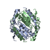
| Title | PRD1 vertex protein P5 | ||||||
 Components Components | Minor capsid protein | ||||||
 Keywords Keywords | VIRAL PROTEIN / beta-spiral / beta-jelly-roll | ||||||
| Function / homology |  Function and homology information Function and homology information | ||||||
| Biological species |   Enterobacteria phage PRD1 (virus) Enterobacteria phage PRD1 (virus) | ||||||
| Method |  X-RAY DIFFRACTION / SYNCHROTRON / MAD / Resolution: 2 Å X-RAY DIFFRACTION / SYNCHROTRON / MAD / Resolution: 2 Å | ||||||
 Authors Authors | Merckel, M.C. / Huiskonen, J.T. / Goldman, A. / Bamford, D.H. / Tuma, R. | ||||||
 Citation Citation | Journal: Mol.Cell / Year: 2005 Title: The structure of the bacteriophage PRD1 spike sheds light on the evolution of viral capsid architecture. Authors: Merckel, M.C. / Huiskonen, J.T. / Bamford, D.H. / Goldman, A. / Tuma, R. | ||||||
| History |
|
- Structure visualization
| Structure viewer | Molecule: MolmilJmol/JSmol |
|---|
- Downloads & links
-Download
| PDBx/mmCIF format | 1yq5.cif.gz | 65.8 KB | Display | PDBx/mmCIF format |
|---|---|---|---|---|
| PDB format | pdb1yq5.ent.gz | 48.8 KB | Display | PDB format |
| PDBx/mmJSON format | 1yq5.json.gz | Tree view | PDBx/mmJSON format | |
| Others |  Other downloads Other downloads |
-Validation report
| Summary document | 1yq5_validation.pdf.gz | 416 KB | Display | wwPDB validaton report |
|---|---|---|---|---|
| Full document | 1yq5_full_validation.pdf.gz | 417.6 KB | Display | |
| Data in XML | 1yq5_validation.xml.gz | 15.1 KB | Display | |
| Data in CIF | 1yq5_validation.cif.gz | 21.3 KB | Display | |
| Arichive directory | https://data.pdbj.org/pub/pdb/validation_reports/yq/1yq5ftp://data.pdbj.org/pub/pdb/validation_reports/yq/1yq5 | HTTPS FTP |
-Related structure data


-Links
 PDBj
PDBj- Assembly
| Deposited unit | 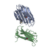
| ||||||||
|---|---|---|---|---|---|---|---|---|---|
| 1 | 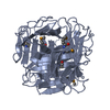
| ||||||||
| 2 | 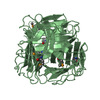
| ||||||||
| Unit cell |
| ||||||||
| Details | The biological assembly is a trimer generated from the chain A in the asymmetric unit by the operations: z,x,y and y,z,x. |
-Components
| #1: Protein | Mass: 14594.293 Da / Num. of mol.: 2 / Fragment: residues 197-340 Source method: isolated from a genetically manipulated source Source: (gene. exp.) Enterobacteria phage PRD1 (virus) / Genus: Tectivirus / Production host:  #2: Water | ChemComp-HOH / |  Mass: 18.015 Da / Num. of mol.: 230 / Source method: isolated from a natural source / Formula: H2O Mass: 18.015 Da / Num. of mol.: 230 / Source method: isolated from a natural source / Formula: H2OHas protein modification | Y | |
|---|
-Experimental details
-Experiment
| Experiment | Method: X-RAY DIFFRACTION / Number of used crystals: 1 |
|---|
- Sample preparation
| Crystal | Density Matthews: 2.97 Å3/Da / Density % sol: 58.7 % |
|---|---|
| Crystal grow | Temperature: 298 K / Method: vapor diffusion, hanging drop / pH: 4.6 Details: Na-formate, Na-acetate, pH 4.6, VAPOR DIFFUSION, HANGING DROP, temperature 298K |
-Data collection
| Diffraction | Mean temperature: 200 K | ||||||||||||
|---|---|---|---|---|---|---|---|---|---|---|---|---|---|
| Diffraction source | Source: SYNCHROTRON / Site: ESRF  / Beamline: BM14 / Wavelength: 0.978335, 0.978497, 0.885595 / Beamline: BM14 / Wavelength: 0.978335, 0.978497, 0.885595 | ||||||||||||
| Detector | Type: MARRESEARCH / Detector: CCD / Date: Oct 1, 2001 | ||||||||||||
| Radiation | Monochromator: Si 111 CHANNEL / Protocol: MAD / Monochromatic (M) / Laue (L): M / Scattering type: x-ray | ||||||||||||
| Radiation wavelength |
| ||||||||||||
| Reflection | Resolution: 2→20 Å / Num. all: 33901 / Num. obs: 33901 / % possible obs: 100 % / Observed criterion σ(F): -3 / Observed criterion σ(I): -3 | ||||||||||||
| Reflection shell | Resolution: 2→2.07 Å / % possible all: 100 |
- Processing
| Software |
| ||||||||||||||||||||
|---|---|---|---|---|---|---|---|---|---|---|---|---|---|---|---|---|---|---|---|---|---|
| Refinement | Method to determine structure: MAD / Resolution: 2→19.91 Å / σ(F): 0 / Stereochemistry target values: Engh & Huber
| ||||||||||||||||||||
| Refinement step | Cycle: LAST / Resolution: 2→19.91 Å
| ||||||||||||||||||||
| Refine LS restraints |
|
