 Movie
Movie Controller

[English] 日本語
 Yorodumi
- PDB-1t0o: The structure of alpha-galactosidase from Trichoderma reesei comp... -
+ Open data

- Basic information
| Entry | Database: PDB / ID: 1t0o | |||||||||
|---|---|---|---|---|---|---|---|---|---|---|
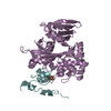
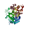
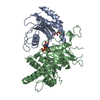
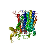
| Title | The structure of alpha-galactosidase from Trichoderma reesei complexed with beta-D-galactose | |||||||||
 Components Components | alpha-galactosidase | |||||||||
 Keywords Keywords | HYDROLASE / (beta/alpha)8 barrel / two domains / glycoprotein / complex / beta-D-galactose | |||||||||
| Function / homology |  Function and homology information Function and homology informationalpha-galactosidase / alpha-galactosidase activity / carbohydrate metabolic process / extracellular region Similarity search - Function | |||||||||
| Biological species |  Hypocrea jecorina (fungus) Hypocrea jecorina (fungus) | |||||||||
| Method |  X-RAY DIFFRACTION / SYNCHROTRON / MOLECULAR REPLACEMENT / Resolution: 1.96 Å X-RAY DIFFRACTION / SYNCHROTRON / MOLECULAR REPLACEMENT / Resolution: 1.96 Å | |||||||||
 Authors Authors | Golubev, A.M. / Nagem, R.A.P. / Brandao Neto, J.R. / Neustroev, K.N. / Eneyskaya, E.V. / Kulminskaya, A.A. / Shabalin, K.A. / Savel'ev, A.N. / Polikarpov, I. | |||||||||
 Citation Citation | Journal: J.Mol.Biol. / Year: 2004 Title: Crystal structure of alpha-galactosidase from Trichoderma reesei and its complex with galactose: implications for catalytic mechanism Authors: Golubev, A.M. / Nagem, R.A.P. / Brandao Neto, J.R. / Neustroev, K.N. / Eneyskaya, E.V. / Kulminskaya, A.A. / Shabalin, K.A. / Savel'ev, A.N. / Polikarpov, I. #1: Journal: J.Mol.Biol. / Year: 1993Title: Crystallization of alpha-galactosidase from Trichoderma reesei. Authors: Golubev, A.M. / Neustroev, K.N. | |||||||||
| History |
| |||||||||
| Remark 999 | SEQUENCE Leu37, Pro70, Ala72, Ala88, Asn148, Phe151, Lys153, Thr161, Asp165, Thr167, Gly185, ...SEQUENCE Leu37, Pro70, Ala72, Ala88, Asn148, Phe151, Lys153, Thr161, Asp165, Thr167, Gly185, His196, Met202, Gln207, Ser216, Asp225, Asn230, Arg236, Leu238, Leu240, Leu245, Asp249, Met258, Asn297, Asn300, Ile327, Val335, Tyr337, Phe341, Val355, Ile360, Ala362, Thr363, Asn369, His378, Ser386, Asp389, Ala398, Gln415, Arg416 differ from the sequence database. These residues were used on the basis of the electron density map. The difference may be due to the use of different strains of Trichoderma reesei for sequencing and for the X-ray structure. |
- Structure visualization
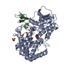
| Structure viewer | Molecule: MolmilJmol/JSmol |
|---|
- Downloads & links
-Download
| PDBx/mmCIF format | 1t0o.cif.gz | 108.8 KB | Display | PDBx/mmCIF format |
|---|---|---|---|---|
| PDB format | pdb1t0o.ent.gz | 81.9 KB | Display | PDB format |
| PDBx/mmJSON format | 1t0o.json.gz | Tree view | PDBx/mmJSON format | |
| Others |  Other downloads Other downloads |
-Validation report
| Summary document | 1t0o_validation.pdf.gz | 1.7 MB | Display | wwPDB validaton report |
|---|---|---|---|---|
| Full document | 1t0o_full_validation.pdf.gz | 1.7 MB | Display | |
| Data in XML | 1t0o_validation.xml.gz | 22.5 KB | Display | |
| Data in CIF | 1t0o_validation.cif.gz | 33.4 KB | Display | |
| Arichive directory | https://data.pdbj.org/pub/pdb/validation_reports/t0/1t0oftp://data.pdbj.org/pub/pdb/validation_reports/t0/1t0o | HTTPS FTP |
-Related structure data
| Related structure data |  1sznSC S: Starting model for refinement C: citing same article ( |
|---|---|
| Similar structure data | |
| Other databases |
|


-Links
 PDBj
PDBj

- Assembly
| Deposited unit | 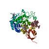
| ||||||||
|---|---|---|---|---|---|---|---|---|---|
| 1 |
| ||||||||
| Unit cell |
| ||||||||
| Details | The biological assembly is a monomer |
-Components
-Protein / Non-polymers , 2 types, 385 molecules A
| #1: Protein | Mass: 45610.863 Da / Num. of mol.: 1 / Source method: isolated from a natural source / Source: (natural) Hypocrea jecorina (fungus)References: GenBank: 1580816, UniProt: Q92456*PLUS, alpha-galactosidase |
|---|---|
| #6: Water | ChemComp-HOH / Mass: 18.015 Da / Num. of mol.: 384 / Source method: isolated from a natural source / Formula: H2O |
-Sugars , 4 types, 5 molecules 
| #2: Polysaccharide | Source method: isolated from a genetically manipulated source #3: Polysaccharide | alpha-D-mannopyranose-(1-3)-[alpha-D-mannopyranose-(1-6)]alpha-D-mannopyranose-(1-6)-[alpha-D- ...alpha-D-mannopyranose-(1-3)-[alpha-D-mannopyranose-(1-6)]alpha-D-mannopyranose-(1-6)-[alpha-D-mannopyranose-(1-3)]beta-D-mannopyranose-(1-4)-2-acetamido-2-deoxy-beta-D-glucopyranose-(1-4)-2-acetamido-2-deoxy-beta-D-glucopyranose | Source method: isolated from a genetically manipulated source #4: Polysaccharide | alpha-D-mannopyranose-(1-3)-beta-D-mannopyranose-(1-4)-2-acetamido-2-deoxy-beta-D-glucopyranose-(1- ...alpha-D-mannopyranose-(1-3)-beta-D-mannopyranose-(1-4)-2-acetamido-2-deoxy-beta-D-glucopyranose-(1-4)-2-acetamido-2-deoxy-beta-D-glucopyranose | Source method: isolated from a genetically manipulated source #5: Sugar | ChemComp-GAL / |  Type: D-saccharide, beta linking / Mass: 180.156 Da / Num. of mol.: 1 Type: D-saccharide, beta linking / Mass: 180.156 Da / Num. of mol.: 1Source method: isolated from a genetically manipulated source Formula: C6H12O6 |
|---|
-Details
| Has protein modification | Y |
|---|
-Experimental details
-Experiment
| Experiment | Method: X-RAY DIFFRACTION / Number of used crystals: 1 |
|---|
- Sample preparation
| Crystal | Density Matthews: 2.7 Å3/Da / Density % sol: 54.07 % |
|---|---|
| Crystal grow | Temperature: 293 K / Method: vapor diffusion, hanging drop / pH: 6 Details: EG 6000, potassium phosphate, sodium phosphate, D-galactose, VAPOR DIFFUSION, HANGING DROP, temperature 293K |
-Data collection
| Diffraction | Mean temperature: 277 K |
|---|---|
| Diffraction source | Source: SYNCHROTRON / Site: LNLS  / Beamline: D03B-MX1 / Wavelength: 1.54 Å / Beamline: D03B-MX1 / Wavelength: 1.54 Å |
| Detector | Type: MARRESEARCH / Detector: IMAGE PLATE / Date: Sep 25, 1998 |
| Radiation | Monochromator: Si 111 CHANNEL / Protocol: SINGLE WAVELENGTH / Monochromatic (M) / Laue (L): M / Scattering type: x-ray |
| Radiation wavelength | Wavelength: 1.54 Å / Relative weight: 1 |
| Reflection | Resolution: 2→9.9 Å / Num. obs: 28741 / % possible obs: 63.6 % / Observed criterion σ(F): 0 / Observed criterion σ(I): 1 / Redundancy: 2.9 % / Biso Wilson estimate: 25.2 Å2 / Rsym value: 0.07 / Net I/σ(I): 10.1 |
| Reflection shell | Resolution: 2→2.05 Å / Rsym value: 0.364 / % possible all: 64 |
- Processing
| Software |
| |||||||||||||||||||||||||||||||||||||||||||||||||||||||||||||||||||||||||||
|---|---|---|---|---|---|---|---|---|---|---|---|---|---|---|---|---|---|---|---|---|---|---|---|---|---|---|---|---|---|---|---|---|---|---|---|---|---|---|---|---|---|---|---|---|---|---|---|---|---|---|---|---|---|---|---|---|---|---|---|---|---|---|---|---|---|---|---|---|---|---|---|---|---|---|---|---|
| Refinement | Method to determine structure: MOLECULAR REPLACEMENT Starting model: PDB ENTRY 1SZN Resolution: 1.96→9 Å / Cor.coef. Fo:Fc: 0.969 / Cor.coef. Fo:Fc free: 0.944 / SU B: 4.034 / SU ML: 0.115 / Cross valid method: THROUGHOUT / σ(F): 0 / σ(I): 0 / ESU R: 0.225 / ESU R Free: 0.184 / Stereochemistry target values: MAXIMUM LIKELIHOOD
| |||||||||||||||||||||||||||||||||||||||||||||||||||||||||||||||||||||||||||
| Solvent computation | Ion probe radii: 0.8 Å / Shrinkage radii: 0.8 Å / VDW probe radii: 1.4 Å / Solvent model: BABINET MODEL WITH MASK | |||||||||||||||||||||||||||||||||||||||||||||||||||||||||||||||||||||||||||
| Displacement parameters | Biso mean: 25.205 Å2
| |||||||||||||||||||||||||||||||||||||||||||||||||||||||||||||||||||||||||||
| Refinement step | Cycle: LAST / Resolution: 1.96→9 Å
| |||||||||||||||||||||||||||||||||||||||||||||||||||||||||||||||||||||||||||
| Refine LS restraints |
| |||||||||||||||||||||||||||||||||||||||||||||||||||||||||||||||||||||||||||
| LS refinement shell | Resolution: 1.96→2.025 Å / Total num. of bins used: 15 /
|
