 Movie
Movie Controller

[English] 日本語
 Yorodumi
- PDB-1l2g: Structure of a C-terminally truncated form of glycoprotein D from... -
+ Open data

- Basic information
| Entry | Database: PDB / ID: 1l2g | ||||||
|---|---|---|---|---|---|---|---|
| Title | Structure of a C-terminally truncated form of glycoprotein D from HSV-1 | ||||||
 Components Components | Glycoprotein D | ||||||
 Keywords Keywords | VIRAL PROTEIN / Ig fold / viral envelope glycoprotein | ||||||
| Function / homology |  Function and homology information Function and homology informationhost cell Golgi apparatus / entry receptor-mediated virion attachment to host cell / viral envelope / symbiont entry into host cell / virion membrane / metal ion binding / membrane Similarity search - Function | ||||||
| Biological species |   Human herpesvirus 1 (Herpes simplex virus type 1) Human herpesvirus 1 (Herpes simplex virus type 1) | ||||||
| Method |  X-RAY DIFFRACTION / SYNCHROTRON / MOLECULAR REPLACEMENT / Resolution: 2.85 Å X-RAY DIFFRACTION / SYNCHROTRON / MOLECULAR REPLACEMENT / Resolution: 2.85 Å | ||||||
 Authors Authors | Carfi, A. / Willis, S.H. / Whitbeck, J.C. / Krummenacher, C. / Cohen, G.H. / Eisenberg, R.J. / Wiley, D.C. | ||||||
 Citation Citation | Journal: Mol.Cell / Year: 2001 Title: Herpes simplex virus glycoprotein D bound to the human receptor HveA. Authors: Carfi, A. / Willis, S.H. / Whitbeck, J.C. / Krummenacher, C. / Cohen, G.H. / Eisenberg, R.J. / Wiley, D.C. #1: Journal: Cell(Cambridge,Mass.) / Year: 1996Title: HERPES SIMPLEX VIRUS-1 ENTRY INTO CELLS MEDIATED BY A NOVEL MEMBER OF THE TNF/NGF RECEPTOR FAMILY Authors: MONTGOMERY, R.I. / WARNER, M.S. / LUM, B.J. / SPEAR, P.G. | ||||||
| History |
| ||||||
| Remark 300 | BIOMOLECULE: 1, 2, 3, 4 ACCORDING TO THE AUTHOR, THIS C-TERMINALLY TRUNCATED GD MOLECULE (RESIDUES ...BIOMOLECULE: 1, 2, 3, 4 ACCORDING TO THE AUTHOR, THIS C-TERMINALLY TRUNCATED GD MOLECULE (RESIDUES 1 TO 285) IS MONOMERIC IN SOLUTION, BUT FORMED DIMERS IN THE CRYSTAL. THIS ENTRY CONTAINS TWO DIMERS, AN AB DIMER CONSISTING OF CHAINS A AND B, AND A CD DIMER CONSISTING OF CHAINS C AND D. |
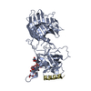
- Structure visualization
| Structure viewer | Molecule: MolmilJmol/JSmol |
|---|
- Downloads & links
-Download
| PDBx/mmCIF format | 1l2g.cif.gz | 375.8 KB | Display | PDBx/mmCIF format |
|---|---|---|---|---|
| PDB format | pdb1l2g.ent.gz | 312.1 KB | Display | PDB format |
| PDBx/mmJSON format | 1l2g.json.gz | Tree view | PDBx/mmJSON format | |
| Others |  Other downloads Other downloads |
-Validation report
| Summary document | 1l2g_validation.pdf.gz | 545.2 KB | Display | wwPDB validaton report |
|---|---|---|---|---|
| Full document | 1l2g_full_validation.pdf.gz | 642.3 KB | Display | |
| Data in XML | 1l2g_validation.xml.gz | 81.3 KB | Display | |
| Data in CIF | 1l2g_validation.cif.gz | 94.5 KB | Display | |
| Arichive directory | https://data.pdbj.org/pub/pdb/validation_reports/l2/1l2gftp://data.pdbj.org/pub/pdb/validation_reports/l2/1l2g | HTTPS FTP |
-Related structure data
| Related structure data | 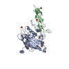 1jmaSC S: Starting model for refinement C: citing same article ( |
|---|---|
| Similar structure data |


-Links
 PDBj
PDBj- Assembly
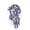
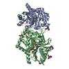
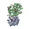
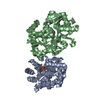
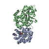
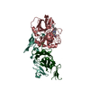
| Deposited unit | 
| ||||||||
|---|---|---|---|---|---|---|---|---|---|
| 1 | 
| ||||||||
| 2 | 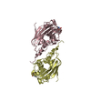
| ||||||||
| 3 | 
| ||||||||
| 4 | 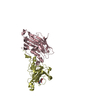
| ||||||||
| Unit cell |
| ||||||||
| Number of models | 2 |
-Components
| #1: Protein | Mass: 31837.168 Da / Num. of mol.: 4 / Fragment: Ectodomain Source method: isolated from a genetically manipulated source Source: (gene. exp.) Human herpesvirus 1 (Herpes simplex virus type 1)Genus: Simplexvirus / Plasmid: PFASTBAC-DUAL / Production host:   Spodoptera frugiperda (fall armyworm) / Strain (production host): Sf9 / References: UniProt: P57083 Spodoptera frugiperda (fall armyworm) / Strain (production host): Sf9 / References: UniProt: P57083#2: Sugar | ChemComp-NAG /   Type: D-saccharide, beta linking / Mass: 221.208 Da / Num. of mol.: 4 Type: D-saccharide, beta linking / Mass: 221.208 Da / Num. of mol.: 4Source method: isolated from a genetically manipulated source Formula: C8H15NO6 Has protein modification | Y | |
|---|
-Experimental details
-Experiment
| Experiment | Method: X-RAY DIFFRACTION / Number of used crystals: 1 |
|---|
- Sample preparation
| Crystal | Density Matthews: 2.82 Å3/Da / Density % sol: 56.38 % |
|---|---|
| Crystal grow | Temperature: 293 K / Method: vapor diffusion, sitting drop / pH: 9 Details: Ammonium Sulfate, pH 9, VAPOR DIFFUSION, SITTING DROP, temperature 293.0K |
-Data collection
| Diffraction | Mean temperature: 100 K |
|---|---|
| Diffraction source | Source: SYNCHROTRON / Site: NSLS  / Beamline: X25 / Wavelength: 1.1 Å / Beamline: X25 / Wavelength: 1.1 Å |
| Detector | Type: BRANDEIS - B4 / Detector: CCD / Date: Oct 10, 1999 |
| Radiation | Monochromator: Si 111 CHANNEL / Protocol: SINGLE WAVELENGTH / Monochromatic (M) / Laue (L): M / Scattering type: x-ray |
| Radiation wavelength | Wavelength: 1.1 Å / Relative weight: 1 |
| Reflection | Resolution: 2.85→30 Å / Num. obs: 29319 / % possible obs: 87.1 % / Observed criterion σ(F): -2 / Observed criterion σ(I): -3 / Redundancy: 2.5 % / Biso Wilson estimate: 51 Å2 / Rsym value: 0.076 / Net I/σ(I): 11 |
| Reflection shell | Resolution: 2.85→2.95 Å / Mean I/σ(I) obs: 2.7 / Rsym value: 0.23 / % possible all: 67.3 |
- Processing
| Software |
| |||||||||||||||||||||||||
|---|---|---|---|---|---|---|---|---|---|---|---|---|---|---|---|---|---|---|---|---|---|---|---|---|---|---|
| Refinement | Method to determine structure: MOLECULAR REPLACEMENT Starting model: PDB ENTRY 1JMA Resolution: 2.85→30 Å / Cross valid method: THROUGHOUT / σ(F): 2 / Stereochemistry target values: Engh & Huber Details: THE CRYSTALS ARE MEROHEDRALLY TWINNED. THE TWINNING OPERATION IS A 2 FOLD ROTATION PARALLEL TO THE A AXIS. THE TWO BLOCKS ARE REPRESENTED IN THIS ENTRY BY TWO MODELS (MODEL 1 AND MODEL 2) ...Details: THE CRYSTALS ARE MEROHEDRALLY TWINNED. THE TWINNING OPERATION IS A 2 FOLD ROTATION PARALLEL TO THE A AXIS. THE TWO BLOCKS ARE REPRESENTED IN THIS ENTRY BY TWO MODELS (MODEL 1 AND MODEL 2) CONTAINING 4 CHAINS (ABCD) EACH. MOLECULES ABCD IN MODEL 1 ARE RELATED BY A 2 FOLD ROTATION AXIS (TWIN OPERATION) TO MOLECULES ABCD OF MODEL 2. NO DETWINNING OF THE DATA WAS ATTEMPTED.
| |||||||||||||||||||||||||
| Displacement parameters |
| |||||||||||||||||||||||||
| Refinement step | Cycle: LAST / Resolution: 2.85→30 Å
| |||||||||||||||||||||||||
| Refine LS restraints |
| |||||||||||||||||||||||||
| LS refinement shell | Resolution: 2.85→2.98 Å
|
