 Movie
Movie Controller

[English] 日本語
 Yorodumi
- PDB-1l1g: The Structure of Porcine Pancreatic Elastase Complexed with Xenon... -
+ Open data

- Basic information
| Entry | Database: PDB / ID: 1l1g | ||||||
|---|---|---|---|---|---|---|---|
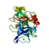
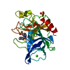
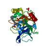
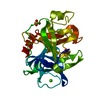
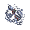
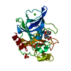
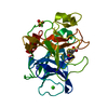
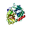
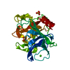
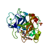
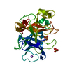
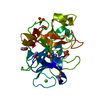
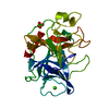
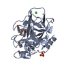
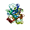
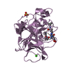
| Title | The Structure of Porcine Pancreatic Elastase Complexed with Xenon and Bromide, Cryoprotected with Glycerol | ||||||
 Components Components | ELASTASE 1 | ||||||
 Keywords Keywords | HYDROLASE / Xenon / Bromide / PPE | ||||||
| Function / homology |  Function and homology information Function and homology informationpancreatic elastase / serine-type endopeptidase activity / proteolysis / extracellular space / metal ion binding Similarity search - Function | ||||||
| Biological species |  | ||||||
| Method |  X-RAY DIFFRACTION / SYNCHROTRON / MAD / Resolution: 1.5 Å X-RAY DIFFRACTION / SYNCHROTRON / MAD / Resolution: 1.5 Å | ||||||
 Authors Authors | Panjikar, S. / Tucker, P.A. | ||||||
 Citation Citation | Journal: Acta Crystallogr.,Sect.D / Year: 2002 Title: Xenon derivatization of halide-soaked protein crystals. Authors: Panjikar, S. / Tucker, P.A. | ||||||
| History |
|
- Structure visualization
| Structure viewer | Molecule: MolmilJmol/JSmol |
|---|
- Downloads & links
-Download
| PDBx/mmCIF format | 1l1g.cif.gz | 65.5 KB | Display | PDBx/mmCIF format |
|---|---|---|---|---|
| PDB format | pdb1l1g.ent.gz | 46.4 KB | Display | PDB format |
| PDBx/mmJSON format | 1l1g.json.gz | Tree view | PDBx/mmJSON format | |
| Others |  Other downloads Other downloads |
-Validation report
| Summary document | 1l1g_validation.pdf.gz | 441.7 KB | Display | wwPDB validaton report |
|---|---|---|---|---|
| Full document | 1l1g_full_validation.pdf.gz | 442.6 KB | Display | |
| Data in XML | 1l1g_validation.xml.gz | 13.6 KB | Display | |
| Data in CIF | 1l1g_validation.cif.gz | 19.5 KB | Display | |
| Arichive directory | https://data.pdbj.org/pub/pdb/validation_reports/l1/1l1gftp://data.pdbj.org/pub/pdb/validation_reports/l1/1l1g | HTTPS FTP |
-Related structure data
| Related structure data | 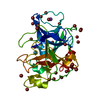 1l0zC 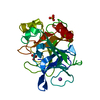 1qnjS S: Starting model for refinement C: citing same article ( |
|---|---|
| Similar structure data |


-Links
 PDBj
PDBj


- Assembly
| Deposited unit | 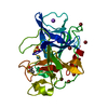
| ||||||||
|---|---|---|---|---|---|---|---|---|---|
| 1 |
| ||||||||
| Unit cell |
|
-Components
-Protein , 1 types, 1 molecules A
| #1: Protein | Mass: 25929.016 Da / Num. of mol.: 1 / Source method: isolated from a natural source / Source: (natural) |
|---|
-Non-polymers , 6 types, 225 molecules 


| #2: Chemical | ChemComp-SO4 /  Mass: 96.063 Da / Num. of mol.: 1 / Source method: obtained synthetically / Formula: SO4 Mass: 96.063 Da / Num. of mol.: 1 / Source method: obtained synthetically / Formula: SO4 | ||||||
|---|---|---|---|---|---|---|---|
| #3: Chemical | ChemComp-NA /  Mass: 22.990 Da / Num. of mol.: 1 / Source method: obtained synthetically / Formula: Na Mass: 22.990 Da / Num. of mol.: 1 / Source method: obtained synthetically / Formula: Na | ||||||
| #4: Chemical | ChemComp-BR /  Mass: 79.904 Da / Num. of mol.: 8 / Source method: obtained synthetically / Formula: Br Mass: 79.904 Da / Num. of mol.: 8 / Source method: obtained synthetically / Formula: Br#5: Chemical | ChemComp-XE / |  Mass: 131.293 Da / Num. of mol.: 1 / Source method: obtained synthetically / Formula: Xe Mass: 131.293 Da / Num. of mol.: 1 / Source method: obtained synthetically / Formula: Xe#6: Chemical | ChemComp-GOL / |  Mass: 92.094 Da / Num. of mol.: 1 / Source method: obtained synthetically / Formula: C3H8O3 Mass: 92.094 Da / Num. of mol.: 1 / Source method: obtained synthetically / Formula: C3H8O3#7: Water | ChemComp-HOH / | Mass: 18.015 Da / Num. of mol.: 213 / Source method: isolated from a natural source / Formula: H2O |
-Details
| Has protein modification | Y |
|---|
-Experimental details
-Experiment
| Experiment | Method: X-RAY DIFFRACTION / Number of used crystals: 1 |
|---|
- Sample preparation
| Crystal | Density Matthews: 2.07 Å3/Da / Density % sol: 40.65 % |
|---|---|
| Crystal grow | Temperature: 298 K / Method: vapor diffusion, hanging drop / pH: 5 Details: 10 mM sodium acetate, pH 5, VAPOR DIFFUSION, HANGING DROP, temperature 298K |
| Crystal grow | *PLUS Details: Shotton, D.M., (1968) J. Mol. Biol., 32, 155. |
-Data collection
| Diffraction | Mean temperature: 100 K |
|---|---|
| Diffraction source | Source: SYNCHROTRON / Site: EMBL/DESY, HAMBURG  / Beamline: X13 / Wavelength: 0.8 Å / Beamline: X13 / Wavelength: 0.8 Å |
| Detector | Type: MARRESEARCH / Detector: CCD / Date: Oct 29, 2001 |
| Radiation | Protocol: SAS / Monochromatic (M) / Laue (L): M / Scattering type: x-ray |
| Radiation wavelength | Wavelength: 0.8 Å / Relative weight: 1 |
| Reflection | Resolution: 1.5→35 Å / Num. all: 34589 / Num. obs: 34589 / % possible obs: 100 % / Observed criterion σ(I): -3 / Redundancy: 15.4 % / Biso Wilson estimate: 12.4 Å2 |
| Reflection | *PLUS Lowest resolution: 35 Å / Num. obs: 66284 / % possible obs: 99 % / Num. measured all: 1017166 / Rmerge(I) obs: 0.076 |
| Reflection shell | *PLUS Highest resolution: 1.2 Å / Lowest resolution: 1.22 Å / % possible obs: 100 % / Redundancy: 10 % / Rmerge(I) obs: 0.457 / Mean I/σ(I) obs: 8 |
- Processing
| Software |
| ||||||||||||||||||||
|---|---|---|---|---|---|---|---|---|---|---|---|---|---|---|---|---|---|---|---|---|---|
| Refinement | Method to determine structure: MAD Starting model: PDB ENTRY 1QNJ Resolution: 1.5→35 Å / Isotropic thermal model: Individual / Cross valid method: THROUGHOUT / σ(F): 0 / Stereochemistry target values: Engh & Huber / Details: HOH 1267 IS AN ALTERNATE CONFORMATION OF BR 267
| ||||||||||||||||||||
| Refinement step | Cycle: LAST / Resolution: 1.5→35 Å
| ||||||||||||||||||||
| Refine LS restraints |
| ||||||||||||||||||||
| Software | *PLUS Name: SHELXL / Version: 97 / Classification: refinement | ||||||||||||||||||||
| Refinement | *PLUS Lowest resolution: 35 Å / Rfactor all: 0.173 | ||||||||||||||||||||
| Solvent computation | *PLUS | ||||||||||||||||||||
| Displacement parameters | *PLUS | ||||||||||||||||||||
| Refine LS restraints | *PLUS
|
