 Movie
Movie Controller

[English] 日本語
 Yorodumi
- EMDB-45563: Cryo-EM structure of myosin-1c bound to F-actin in the ADP-A state -
+ Open data

- Basic information
| Entry |  | |||||||||
|---|---|---|---|---|---|---|---|---|---|---|
| Title | Cryo-EM structure of myosin-1c bound to F-actin in the ADP-A state | |||||||||
 Map data Map data | ||||||||||
 Sample Sample |
| |||||||||
 Keywords Keywords | F-actin / myosin / myosin-1c / cellular motility / cryo-EM / actomyosin. / STRUCTURAL PROTEIN / MOTOR PROTEIN | |||||||||
| Function / homology |  Function and homology information Function and homology informationpositive regulation of cellular response to insulin stimulus / stereocilium membrane / B-WICH complex positively regulates rRNA expression / CaMK IV-mediated phosphorylation of CREB / Cam-PDE 1 activation / CREB1 phosphorylation through the activation of CaMKII/CaMKK/CaMKIV cascasde / Glycogen breakdown (glycogenolysis) / Activation of RAC1 downstream of NMDARs / Sodium/Calcium exchangers / Activation of Ca-permeable Kainate Receptor ...positive regulation of cellular response to insulin stimulus / stereocilium membrane / B-WICH complex positively regulates rRNA expression / CaMK IV-mediated phosphorylation of CREB / Cam-PDE 1 activation / CREB1 phosphorylation through the activation of CaMKII/CaMKK/CaMKIV cascasde / Glycogen breakdown (glycogenolysis) / Activation of RAC1 downstream of NMDARs / Sodium/Calcium exchangers / Activation of Ca-permeable Kainate Receptor / Synthesis of IP3 and IP4 in the cytosol / CLEC7A (Dectin-1) induces NFAT activation / RHO GTPases activate PAKs / Calmodulin induced events / Inactivation, recovery and regulation of the phototransduction cascade / Tetrahydrobiopterin (BH4) synthesis, recycling, salvage and regulation / eNOS activation / Reduction of cytosolic Ca++ levels / Unblocking of NMDA receptors, glutamate binding and activation / Protein methylation / Calcineurin activates NFAT / Ion transport by P-type ATPases / RAF activation / VEGFR2 mediated vascular permeability / RAS processing / vesicle transport along actin filament / Ca2+ pathway / RHO GTPases activate IQGAPs / Extra-nuclear estrogen signaling / FCERI mediated Ca+2 mobilization / RAF/MAP kinase cascade / PKA activation / Smooth Muscle Contraction / Regulation of actin dynamics for phagocytic cup formation / Platelet degranulation / High laminar flow shear stress activates signaling by PIEZO1 and PECAM1:CDH5:KDR in endothelial cells / Stimuli-sensing channels / B-WICH complex / stereocilium / establishment of protein localization to mitochondrial membrane / Ion homeostasis / type 3 metabotropic glutamate receptor binding / myosin complex / protein targeting to membrane / vascular endothelial growth factor signaling pathway / regulation of bicellular tight junction assembly / positive regulation of transcription by RNA polymerase III / cytoskeletal motor activator activity / response to corticosterone / negative regulation of ryanodine-sensitive calcium-release channel activity / organelle localization by membrane tethering / myosin heavy chain binding / microfilament motor activity / mitochondrion-endoplasmic reticulum membrane tethering / autophagosome membrane docking / tropomyosin binding / regulation of cardiac muscle cell action potential / presynaptic endocytosis / actin filament bundle / nitric-oxide synthase binding / regulation of synaptic vesicle exocytosis / troponin I binding / filamentous actin / microvillus / positive regulation of transcription by RNA polymerase I / mesenchyme migration / calcineurin-mediated signaling / brush border / actin filament bundle assembly / skeletal muscle myofibril / adenylate cyclase binding / striated muscle thin filament / regulation of ryanodine-sensitive calcium-release channel activity / protein phosphatase activator activity / skeletal muscle thin filament assembly / actin monomer binding / lateral plasma membrane / catalytic complex / positive regulation of protein targeting to membrane / detection of calcium ion / regulation of synaptic vesicle endocytosis / regulation of cardiac muscle contraction / postsynaptic cytosol / cellular response to interferon-beta / phosphatidylinositol 3-kinase binding / calcium channel inhibitor activity / presynaptic cytosol / skeletal muscle fiber development / regulation of release of sequestered calcium ion into cytosol by sarcoplasmic reticulum / stress fiber / phagocytic vesicle / titin binding / sperm midpiece / actin filament polymerization / voltage-gated potassium channel complex / calcium channel complex / regulation of heart rate / basal plasma membrane / cytoplasmic vesicle membrane / calyx of Held Similarity search - Function | |||||||||
| Biological species |  | |||||||||
| Method | helical reconstruction / cryo EM / Resolution: 2.8 Å | |||||||||
 Authors Authors | Chavali SS / Sindelar CV / Ostap ME | |||||||||
| Funding support |  United States, 2 items United States, 2 items
| |||||||||
 Citation Citation | Journal: Proc Natl Acad Sci U S A / Year: 2025 Title: High-resolution structures of Myosin-IC reveal a unique actin-binding orientation, ADP release pathway, and power stroke trajectory. Authors: Sai Shashank Chavali / Peter J Carman / Henry Shuman / E Michael Ostap / Charles V Sindelar / Abstract: Myosin-IC (myo1c) is a class-I myosin that supports transport and remodeling of the plasma membrane and membrane-bound vesicles. Like other members of the myosin family, its biochemical kinetics are ...Myosin-IC (myo1c) is a class-I myosin that supports transport and remodeling of the plasma membrane and membrane-bound vesicles. Like other members of the myosin family, its biochemical kinetics are altered in response to changes in mechanical loads that resist the power stroke. However, myo1c is unique in that the primary force-sensitive kinetic transition is the isomerization that follows ATP binding, not ADP release as in other slow myosins. Myo1c also powers actin gliding along curved paths, propelling actin filaments in leftward circles. To understand the origins of this unique force-sensing and motile behavior, we solved actin-bound myo1c cryo-EM structures in the presence and absence of ADP. Our structures reveal that in contrast with other myosins, the myo1c lever arm swing is skewed, partly due to a different actin interface that reorients the motor domain on actin. The structures also reveal unique nucleotide-dependent behavior of both the nucleotide pocket as well as an element called the N-terminal extension (NTE). We incorporate these observations into a model that explains why force primarily regulates ATP binding in myo1c, rather than ADP release as in other myosins. Integrating our cryo-EM data with available crystallography structures allows the modeling of full-length myo1c during force generation, supplying insights into its role in membrane remodeling. These results highlight how relatively minor sequence differences in members of the myosin superfamily can significantly alter power stroke geometry and force-sensing properties, with important implications for biological function. | |||||||||
| History |
|
- Structure visualization
| Supplemental images |
|---|

- Downloads & links
-EMDB archive
| Map data | emd_45563.map.gz | 1.7 MB | EMDB map data format | |
|---|---|---|---|---|
| Header (meta data) | emd-45563-v30.xmlemd-45563.xml | 19.4 KB 19.4 KB | Display Display | EMDB header |
| FSC (resolution estimation) | emd_45563_fsc.xml | 9 KB | Display | FSC data file |
| Images |  emd_45563.png emd_45563.png | 53.3 KB | ||
| Masks | emd_45563_msk_1.map | 64 MB | Mask map | |
| Filedesc metadata | emd-45563.cif.gz | 7.1 KB | ||
| Others | emd_45563_half_map_1.map.gzemd_45563_half_map_2.map.gz | 200.4 MB 200.4 MB | ||
| Archive directory |  http://ftp.pdbj.org/pub/emdb/structures/EMD-45563ftp://ftp.pdbj.org/pub/emdb/structures/EMD-45563 http://ftp.pdbj.org/pub/emdb/structures/EMD-45563ftp://ftp.pdbj.org/pub/emdb/structures/EMD-45563 | HTTPS FTP |
-Validation report
| Summary document | emd_45563_validation.pdf.gz | 675.1 KB | Display | EMDB validaton report |
|---|---|---|---|---|
| Full document | emd_45563_full_validation.pdf.gz | 674.7 KB | Display | |
| Data in XML | emd_45563_validation.xml.gz | 19.7 KB | Display | |
| Data in CIF | emd_45563_validation.cif.gz | 25.2 KB | Display | |
| Arichive directory | https://ftp.pdbj.org/pub/emdb/validation_reports/EMD-45563ftp://ftp.pdbj.org/pub/emdb/validation_reports/EMD-45563 | HTTPS FTP |
-Related structure data
| Related structure data |  9cfuMC  9cfvC  9cfwC  9cfxC M: atomic model generated by this map C: citing same article ( |
|---|---|
| Similar structure data |


-Links
| EMDB pages | EMDB (EBI/PDBe) / EMDataResource |
|---|---|
| Related items in Molecule of the Month |


-Map
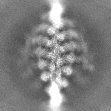
| File | Download / File: emd_45563.map.gz / Format: CCP4 / Size: 216 MB / Type: IMAGE STORED AS FLOATING POINT NUMBER (4 BYTES) | ||||||||||||||||||||||||||||||||||||
|---|---|---|---|---|---|---|---|---|---|---|---|---|---|---|---|---|---|---|---|---|---|---|---|---|---|---|---|---|---|---|---|---|---|---|---|---|---|
| Projections & slices | Image control
Images are generated by Spider. | ||||||||||||||||||||||||||||||||||||
| Voxel size | X=Y=Z: 1.346 Å | ||||||||||||||||||||||||||||||||||||
| Density |
| ||||||||||||||||||||||||||||||||||||
| Symmetry | Space group: 1 | ||||||||||||||||||||||||||||||||||||
| Details | EMDB XML:
|
 Z (Sec.)
 Y (Row.)
 X (Col.)


-Supplemental data
-Mask #1
| File | emd_45563_msk_1.map | ||||||||||||
|---|---|---|---|---|---|---|---|---|---|---|---|---|---|
| Projections & Slices |
| ||||||||||||
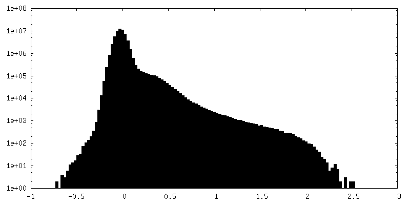
| Density Histograms |


-Half map: #1
| File | emd_45563_half_map_1.map | ||||||||||||
|---|---|---|---|---|---|---|---|---|---|---|---|---|---|
| Projections & Slices |
| ||||||||||||
| Density Histograms |


-Half map: #2
| File | emd_45563_half_map_2.map | ||||||||||||
|---|---|---|---|---|---|---|---|---|---|---|---|---|---|
| Projections & Slices |
| ||||||||||||
| Density Histograms |


- Sample components
-Entire : Complex of myosin-1c with F-actin
| Entire | Name: Complex of myosin-1c with F-actin |
|---|---|
| Components |
|
-Supramolecule #1: Complex of myosin-1c with F-actin
| Supramolecule | Name: Complex of myosin-1c with F-actin / type: complex / ID: 1 / Parent: 0 / Macromolecule list: #1-#3 |
|---|---|
| Source (natural) | Organism: |
| Molecular weight | Theoretical: 220 KDa |
-Macromolecule #1: Actin, alpha skeletal muscle
| Macromolecule | Name: Actin, alpha skeletal muscle / type: protein_or_peptide / ID: 1 / Number of copies: 3 / Enantiomer: LEVO EC number: Hydrolases; Acting on acid anhydrides; Acting on acid anhydrides to facilitate cellular and subcellular movement |
|---|---|
| Source (natural) | Organism: |
| Molecular weight | Theoretical: 41.862613 KDa |
| Sequence | String: DEDETTALVC DNGSGLVKAG FAGDDAPRAV FPSIVGRPRH QGVMVGMGQK DSYVGDEAQS KRGILTLKYP IEHGIITNWD DMEKIWHHT FYNELRVAPE EHPTLLTEAP LNPKANREKM TQIMFETFNV PAMYVAIQAV LSLYASGRTT GIVLDSGDGV T HNVPIYEG ...String: DEDETTALVC DNGSGLVKAG FAGDDAPRAV FPSIVGRPRH QGVMVGMGQK DSYVGDEAQS KRGILTLKYP IEHGIITNWD DMEKIWHHT FYNELRVAPE EHPTLLTEAP LNPKANREKM TQIMFETFNV PAMYVAIQAV LSLYASGRTT GIVLDSGDGV T HNVPIYEG YALPHAIMRL DLAGRDLTDY LMKILTERGY SFVTTAEREI VRDIKEKLCY VALDFENEMA TAASSSSLEK SY ELPDGQV ITIGNERFRC PETLFQPSFI GMESAGIHET TYNSIMKCDI DIRKDLYANN VMSGGTTMYP GIADRMQKEI TAL APSTMK IKIIAPPERK YSVWIGGSIL ASLSTFQQMW ITKQEYDEAG PSIVHRKCF UniProtKB: Actin, alpha skeletal muscle |
-Macromolecule #2: Unconventional myosin-Ic
| Macromolecule | Name: Unconventional myosin-Ic / type: protein_or_peptide / ID: 2 / Number of copies: 1 / Enantiomer: LEVO |
|---|---|
| Source (natural) | Organism: |
| Molecular weight | Theoretical: 92.064203 KDa |
| Recombinant expression | Organism:  unidentified baculovirus unidentified baculovirus |
| Sequence | String: MESALTARDR VGVQDFVLLE NFTSEAAFIE NLRRRFRENL IYTYIGPVLV SVNPYRDLQI YSRQHMERYR GVSFYEVPPH LFAVADTVY RALRTERRDQ AVMISGESGA GKTEATKRLL QFYAETCPAP ERGGAVRDRL LQSNPVLEAF GNAKTLRNDN S SRFGKYMD ...String: MESALTARDR VGVQDFVLLE NFTSEAAFIE NLRRRFRENL IYTYIGPVLV SVNPYRDLQI YSRQHMERYR GVSFYEVPPH LFAVADTVY RALRTERRDQ AVMISGESGA GKTEATKRLL QFYAETCPAP ERGGAVRDRL LQSNPVLEAF GNAKTLRNDN S SRFGKYMD VQFDFKGAPV GGHILSYLLE KSRVVHQNHG ERNFHVFYQL LEGGEEETLR RLGLERNPQS YLYLVKGQCA KV SSINDKS DWKVMRKALS VIDFTEDEVE DLLSIVASVL HLGNIHFAAD EDSNAQVTTE NQLKYLTRLL GVEGTTLREA LTH RKIIAK GEELLSPLNL EQAAYARDAL AKAVYSRTFT WLVRKINRSL ASKDAESPSW RSTTVLGLLD IYGFEVFQHN SFEQ FCINY CNEKLQQLFI ELTLKSEQEE YEAEGIAWEP VQYFNNKIIC DLVEEKFKGI ISILDEECLR PGEATDLTFL EKLED TVKP HPHFLTHKLA DQKTRKSLDR GEFRLLHYAG EVTYSVTGFL DKNNDLLFRN LKETMCSSMN PIMAQCFDKS ELSDKK RPE TVATQFKMSL LQLVEILRSK EPAYIRCIKP NDAKQPGRFD EVLIRHQVKY LGLMENLRVR RAGFAYRRKY EAFLQRY KS LCPETWPMWA GRPQDGVAVL VRHLGYKPEE YKMGRTKIFI RFPKTLFATE DSLEVRRQSL ATKIQAAWRG FHWRQKFL R VKRSAICIQS WWRGTLGRRK AAKRKWAAQT IRRLIRGFIL RHSPRCGGLN DIFEAQKIEW HEAADYKDDD DK UniProtKB: Unconventional myosin-Ic |
-Macromolecule #3: Calmodulin-1
| Macromolecule | Name: Calmodulin-1 / type: protein_or_peptide / ID: 3 / Number of copies: 1 / Enantiomer: LEVO |
|---|---|
| Source (natural) | Organism: |
| Molecular weight | Theoretical: 16.723365 KDa |
| Recombinant expression | Organism: unidentified baculovirus |
| Sequence | String: MADQLTEEQI AEFKEAFSLF DKDGDGTITT KELGTVMRSL GQNPTEAELQ DMINEVDADG NGTIDFPEFL TMMARKMKDT DSEEEIREA FRVFDKDGNG YISAAELRHV MTNLGEKLTD EEVDEMIREA DIDGDGQVNY EEFVQMMTA UniProtKB: Calmodulin-1 |
-Macromolecule #4: ADENOSINE-5'-DIPHOSPHATE
| Macromolecule | Name: ADENOSINE-5'-DIPHOSPHATE / type: ligand / ID: 4 / Number of copies: 4 / Formula: ADP |
|---|---|
| Molecular weight | Theoretical: 427.201 Da |
| Chemical component information |  ChemComp-ADP: |
-Macromolecule #5: MAGNESIUM ION
| Macromolecule | Name: MAGNESIUM ION / type: ligand / ID: 5 / Number of copies: 4 / Formula: MG |
|---|---|
| Molecular weight | Theoretical: 24.305 Da |
-Experimental details
-Structure determination
| Method | cryo EM |
|---|---|
 Processing Processing | helical reconstruction |
| Aggregation state | filament |
-Sample preparation
| Buffer | pH: 7 |
|---|---|
| Vitrification | Cryogen name: ETHANE |
- Electron microscopy
| Microscope | TFS KRIOS |
|---|---|
| Image recording | Film or detector model: GATAN K3 (6k x 4k) / Average electron dose: 52.0 e/Å2 |
| Electron beam | Acceleration voltage: 300 kV / Electron source:  FIELD EMISSION GUN FIELD EMISSION GUN |
| Electron optics | Illumination mode: OTHER / Imaging mode: BRIGHT FIELD / Nominal defocus max: 2.5 µm / Nominal defocus min: 1.2 µm |
| Experimental equipment |  Model: Titan Krios / Image courtesy: FEI Company |
-Image processing
| Final reconstruction | Applied symmetry - Helical parameters - Δz: 28.266 Å Applied symmetry - Helical parameters - Δ&Phi: -167.687 ° Applied symmetry - Helical parameters - Axial symmetry: C1 (asymmetric) Resolution.type: BY AUTHOR / Resolution: 2.8 Å / Resolution method: FSC 0.143 CUT-OFF / Number images used: 671620 |
|---|---|
| Startup model | Type of model: INSILICO MODEL |
| Final angle assignment | Type: NOT APPLICABLE |
| FSC plot (resolution estimation) | 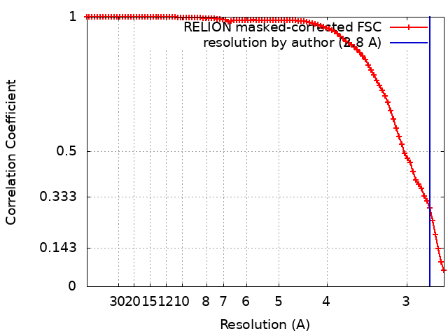 |
