National Institutes of Health/National Institute Of Allergy and Infectious Diseases (NIH/NIAID)
AI144022
米国
引用
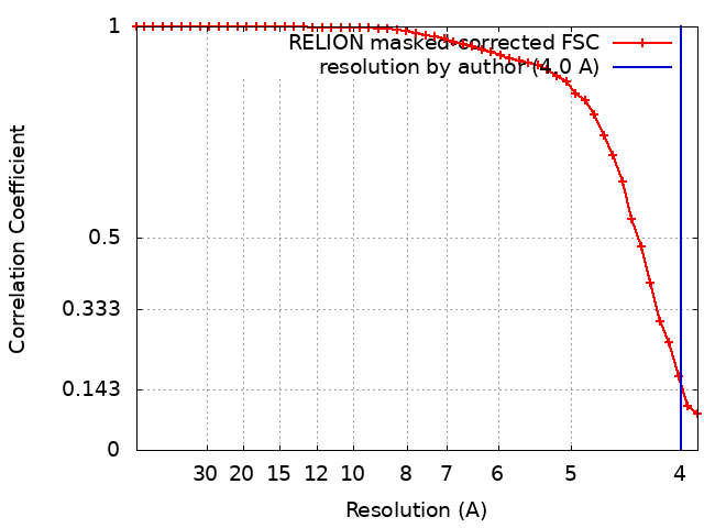
ジャーナル: J Biol Chem / 年: 2023 タイトル: Structure of the Hepatitis B virus capsid quasi-6-fold with a trapped C-terminal domain reveals capsid movements associated with domain exit. 著者: Christine Kim / Christopher J Schlicksup / Carolina Pérez-Segura / Jodi A Hadden-Perilla / Joseph Che-Yen Wang / Adam Zlotnick / 要旨: Many viruses undergo transient conformational change to surveil their environments for receptors and host factors. In Hepatitis B virus (HBV) infection, after the virus enters the cell, it is ...Many viruses undergo transient conformational change to surveil their environments for receptors and host factors. In Hepatitis B virus (HBV) infection, after the virus enters the cell, it is transported to the nucleus by interaction of the HBV capsid with an importin α/β complex. The interaction between virus and importins is mediated by nuclear localization signals on the capsid protein's C-terminal domain (CTD). However, CTDs are located inside the capsid. In this study, we asked where does a CTD exit the capsid, are all quasi-equivalent CTDs created equal, and does the capsid structure deform to facilitate CTD egress from the capsid? Here, we used Impβ as a tool to trap transiently exposed CTDs and examined this complex by cryo-electron microscopy. We examined an asymmetric reconstruction of a T = 4 icosahedral capsid and a focused reconstruction of a quasi-6-fold vertex (3.8 and 4.0 Å resolution, respectively). Both approaches showed that a subset of CTDs extended through a pore in the center of the quasi-6-fold complex. CTD egress was accompanied by enlargement of the pore and subtle changes in quaternary and tertiary structure of the quasi-6-fold. When compared to molecular dynamics simulations, structural changes were within the normal range of capsid flexibility. Although pore diameter was enlarged in the Impβ-bound reconstruction, simulations indicate that CTD egress does not exclusively depend on enlarged pores. In summary, we find that HBV surveillance of its environment by transient exposure of its CTD requires only modest conformational change of the capsid.
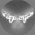
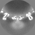
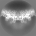
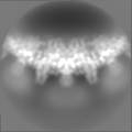
ソフトウェア - 名称: RELION 詳細: Symmetry expansion and alignment was applied to the refined I2 map for focused reconstruction. 1,730,160 quasi-6-fold vertices (referred as hexamers) were extracted. The particles were used ...詳細: Symmetry expansion and alignment was applied to the refined I2 map for focused reconstruction. 1,730,160 quasi-6-fold vertices (referred as hexamers) were extracted. The particles were used to generate a new initial hexamer model de novo with C1 symmetry, which was used as a reference map for subsequent 3D classification.
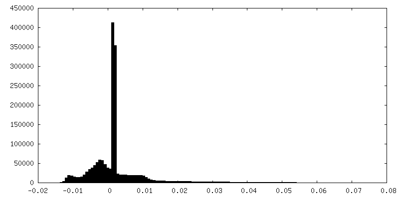
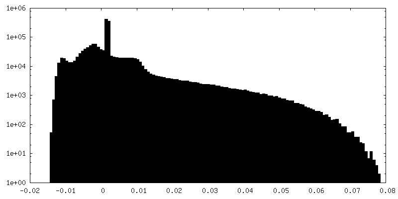
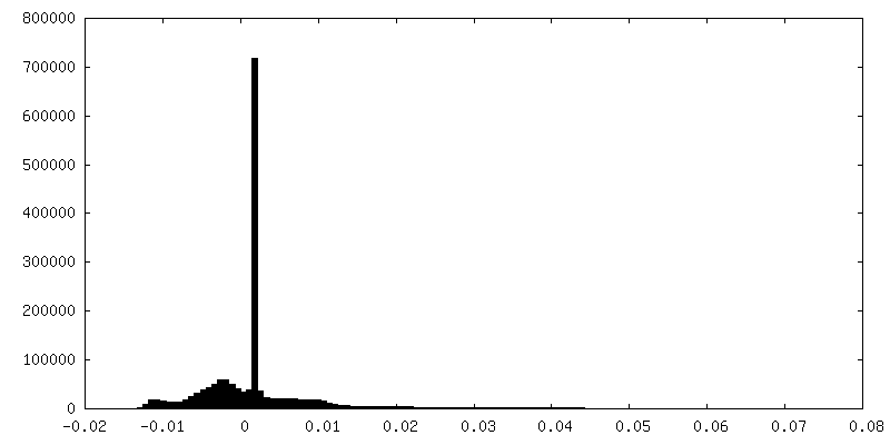
FSC曲線 (解像度の算出)
-
原子モデル構築 1
詳細
A hexamer model was flexibly fitted into the hexamer maps using Alphafold, ISOLDE, PHENIX, and Coot.
精密化
プロトコル: RIGID BODY FIT
得られたモデル
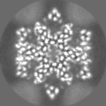
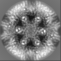
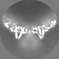
PDB-8ghs: Empty HBV Cp183 capsid with importin-beta, subparticle reconstruction at 2-fold location
 ムービー
ムービー コントローラー


 データを開く
 基本情報
基本情報
 マップデータ
 試料
試料 キーワード
キーワード 機能・相同性情報
 Hepatitis B virus (B 型肝炎ウイルス)
 データ登録者
 米国, 1件
  引用
引用 構造の表示
構造の表示
 ダウンロードとリンク
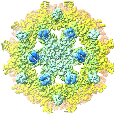 emd_40049.png
 http://ftp.pdbj.org/pub/emdb/structures/EMD-40049


 Z
Z Y
Y X
X


 試料の構成要素
 解析
解析 電子顕微鏡法
 FIELD EMISSION GUN