ムービー
ムービー コントローラー


+ データを開く

- 基本情報
基本情報
| 登録情報 | データベース: EMDB / ID: EMD-3082 | |||||||||
|---|---|---|---|---|---|---|---|---|---|---|
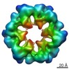
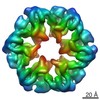
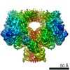
| タイトル | Architecture and nucleotide-driven conformational states of the Rvb1/Rvb2 dodecamer | |||||||||
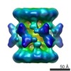
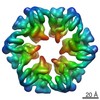
 マップデータ マップデータ | ADP-state Rvb1/2 dodecamer | |||||||||
 試料 試料 |
| |||||||||
 キーワード キーワード | Rv1 / Rvb2 | |||||||||
| 機能・相同性 |  機能・相同性情報 機能・相同性情報TTT Hsp90 cochaperone complex / R2TP complex / Swr1 complex / Ino80 complex / box C/D snoRNP assembly / 3'-5' DNA helicase activity / NuA4 histone acetyltransferase complex / DNA helicase activity / rRNA processing / 5'-3' DNA helicase activity ...TTT Hsp90 cochaperone complex / R2TP complex / Swr1 complex / Ino80 complex / box C/D snoRNP assembly / 3'-5' DNA helicase activity / NuA4 histone acetyltransferase complex / DNA helicase activity / rRNA processing / 5'-3' DNA helicase activity / DNA helicase / protein stabilization / chromatin remodeling / DNA repair / regulation of DNA-templated transcription / regulation of transcription by RNA polymerase II / chromatin / ATP hydrolysis activity / ATP binding / nucleus 類似検索 - 分子機能 | |||||||||
| 生物種 |  | |||||||||
| 手法 | 単粒子再構成法 / クライオ電子顕微鏡法 / 解像度: 7.8 Å | |||||||||
 データ登録者 データ登録者 | Ewens CA / Su M / Zhao L / Houry WA / Southworth DR | |||||||||
 引用 引用 | ジャーナル: Structure / 年: 2016 タイトル: Architecture and Nucleotide-Dependent Conformational Changes of the Rvb1-Rvb2 AAA+ Complex Revealed by Cryoelectron Microscopy. 著者: Caroline A Ewens / Min Su / Liang Zhao / Nardin Nano / Walid A Houry / Daniel R Southworth /   要旨: Rvb1 and Rvb2 are essential AAA+ proteins that interact together during the assembly and activity of diverse macromolecules including chromatin remodelers INO80 and SWR-C, and ribonucleoprotein ...Rvb1 and Rvb2 are essential AAA+ proteins that interact together during the assembly and activity of diverse macromolecules including chromatin remodelers INO80 and SWR-C, and ribonucleoprotein complexes including telomerase and snoRNPs. ATP hydrolysis by Rvb1/2 is required for function; however, the mechanism that drives substrate remodeling is unknown. Here we determined the architecture of the yeast Rvb1/2 dodecamer using cryoelectron microscopy and identify that the substrate-binding insertion domain undergoes conformational changes in response to nucleotide state. 2D and 3D classification defines the dodecamer flexibility, revealing distinct arrangements and the hexamer-hexamer interaction interface. Reconstructions of the apo, ATP, and ADP states identify that Rvb1/2 undergoes substantial conformational changes that include a twist in the insertion-domain position and a corresponding rotation of the AAA+ ring. These results reveal how the ATP hydrolysis cycle of the AAA+ domains directs insertion-domain movements that could provide mechanical force during remodeling or helicase activities. | |||||||||
| 履歴 |
|
- 構造の表示
構造の表示
| ムービー |
ムービービューア |
|---|---|
| 構造ビューア | EMマップ: SurfViewMolmilJmol/JSmol |
| 添付画像 |

- ダウンロードとリンク
-EMDBアーカイブ
| マップデータ | emd_3082.map.gz | 5.3 MB | EMDBマップデータ形式 | |
|---|---|---|---|---|
| ヘッダ (付随情報) | emd-3082-v30.xmlemd-3082.xml | 10.6 KB 10.6 KB | 表示 表示 | EMDBヘッダ |
| 画像 | 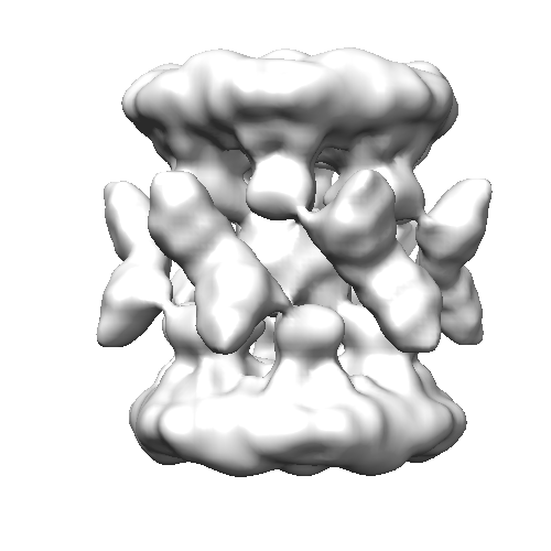 emd_3082.png emd_3082.png | 73.5 KB | ||
| アーカイブディレクトリ |  http://ftp.pdbj.org/pub/emdb/structures/EMD-3082ftp://ftp.pdbj.org/pub/emdb/structures/EMD-3082 http://ftp.pdbj.org/pub/emdb/structures/EMD-3082ftp://ftp.pdbj.org/pub/emdb/structures/EMD-3082 | HTTPS FTP |
-検証レポート
| 文書・要旨 | emd_3082_validation.pdf.gz | 220.7 KB | 表示 | EMDB検証レポート |
|---|---|---|---|---|
| 文書・詳細版 | emd_3082_full_validation.pdf.gz | 219.9 KB | 表示 | |
| XML形式データ | emd_3082_validation.xml.gz | 4.7 KB | 表示 | |
| アーカイブディレクトリ | https://ftp.pdbj.org/pub/emdb/validation_reports/EMD-3082ftp://ftp.pdbj.org/pub/emdb/validation_reports/EMD-3082 | HTTPS FTP |
-関連構造データ


-リンク
| EMDBのページ | EMDB (EBI/PDBe) / EMDataResource |
|---|
-マップ
| ファイル | ダウンロード / ファイル: emd_3082.map.gz / 形式: CCP4 / 大きさ: 7.8 MB / タイプ: IMAGE STORED AS FLOATING POINT NUMBER (4 BYTES) | ||||||||||||||||||||||||||||||||||||||||||||||||||||||||||||
|---|---|---|---|---|---|---|---|---|---|---|---|---|---|---|---|---|---|---|---|---|---|---|---|---|---|---|---|---|---|---|---|---|---|---|---|---|---|---|---|---|---|---|---|---|---|---|---|---|---|---|---|---|---|---|---|---|---|---|---|---|---|
| 注釈 | ADP-state Rvb1/2 dodecamer | ||||||||||||||||||||||||||||||||||||||||||||||||||||||||||||
| 投影像・断面図 | 画像のコントロール
画像は Spider により作成 | ||||||||||||||||||||||||||||||||||||||||||||||||||||||||||||
| ボクセルのサイズ | X=Y=Z: 2 Å | ||||||||||||||||||||||||||||||||||||||||||||||||||||||||||||
| 密度 |
| ||||||||||||||||||||||||||||||||||||||||||||||||||||||||||||
| 対称性 | 空間群: 1 | ||||||||||||||||||||||||||||||||||||||||||||||||||||||||||||
| 詳細 | EMDB XML:
CCP4マップ ヘッダ情報:
| ||||||||||||||||||||||||||||||||||||||||||||||||||||||||||||
 Z (Sec.)
 Y (Row.)
 X (Col.)


-添付データ
- 試料の構成要素
-全体 : ADP-state Rvb1/2 dodecamer
| 全体 | 名称: ADP-state Rvb1/2 dodecamer |
|---|---|
| 要素 |
|
-超分子 #1000: ADP-state Rvb1/2 dodecamer
| 超分子 | 名称: ADP-state Rvb1/2 dodecamer / タイプ: sample / ID: 1000 / 集合状態: heterododecamer / Number unique components: 2 |
|---|---|
| 分子量 | 実験値: 600 KDa / 理論値: 600 KDa 手法: size exclusion chromatography with multiangle light scattering |
-分子 #1: Rvb1
| 分子 | 名称: Rvb1 / タイプ: protein_or_peptide / ID: 1 / Name.synonym: Pontin / コピー数: 6 / 集合状態: Hexamer / 組換発現: Yes |
|---|---|
| 由来(天然) | 生物種: |
| 分子量 | 実験値: 50 KDa / 理論値: 50 KDa |
| 組換発現 | 生物種:  |
| 配列 | UniProtKB: RuvB-like protein 1 |
-分子 #2: Rvb2
| 分子 | 名称: Rvb2 / タイプ: protein_or_peptide / ID: 2 / Name.synonym: Reptin / コピー数: 6 / 集合状態: Hexamer / 組換発現: Yes |
|---|---|
| 由来(天然) | 生物種: |
| 分子量 | 実験値: 50 KDa / 理論値: 50 KDa |
| 組換発現 | 生物種: |
| 配列 | UniProtKB: RuvB-like protein 2 |
-実験情報
-構造解析
| 手法 | クライオ電子顕微鏡法 |
|---|---|
 解析 解析 | 単粒子再構成法 |
| 試料の集合状態 | particle |
-試料調製
| 濃度 | 0.3 mg/mL |
|---|---|
| 緩衝液 | pH: 7.4 詳細: 25mM HEPES, 150mM KCl, 6mM betaME, 5mM MgCl2, 200microM ADP |
| 凍結 | 凍結剤: ETHANE / チャンバー内湿度: 90 % / 装置: FEI VITROBOT MARK IV / 手法: 1s blot |
- 電子顕微鏡法
| 顕微鏡 | FEI TITAN KRIOS |
|---|---|
| 日付 | 2014年11月24日 |
| 撮影 | カテゴリ: CCD / フィルム・検出器のモデル: GATAN K2 (4k x 4k) / 実像数: 3303 / 平均電子線量: 5 e/Å2 詳細: Every image is the average of 30 frames recorded by the direct electron detector |
| 電子線 | 加速電圧: 300 kV / 電子線源:  FIELD EMISSION GUN FIELD EMISSION GUN |
| 電子光学系 | 照射モード: FLOOD BEAM / 撮影モード: DARK FIELD / Cs: 2 mm / 最大 デフォーカス(公称値): 3.0 µm / 最小 デフォーカス(公称値): 1.8 µm / 倍率(公称値): 29000 |
| 試料ステージ | 試料ホルダーモデル: FEI TITAN KRIOS AUTOGRID HOLDER |
| 実験機器 |  モデル: Titan Krios / 画像提供: FEI Company |
-画像解析
| CTF補正 | 詳細: per micrograph |
|---|---|
| 最終 再構成 | 想定した対称性 - 点群: D6 (2回x6回 2面回転対称) アルゴリズム: OTHER / 解像度のタイプ: BY AUTHOR / 解像度: 7.8 Å / 解像度の算出法: OTHER ソフトウェア - 名称: relion, boxer, eman2, cftfind, spider 使用した粒子像数: 34173 |
-原子モデル構築 1
| 初期モデル | PDB ID: Chain - #0 - Chain ID: A / Chain - #1 - Chain ID: B |
|---|---|
| ソフトウェア | 名称: chimera, situs |
| 詳細 | The domains were separately fitted by manual docking |
| 精密化 | 空間: REAL / プロトコル: RIGID BODY FIT / 当てはまり具合の基準: cross correlation |