 Movie
Movie Controller


+ Open data

- Basic information
| Entry | Database: EMDB / ID: EMD-20549 | |||||||||
|---|---|---|---|---|---|---|---|---|---|---|
| Title | Outer Hair Cell, Lateral Wall | |||||||||
 Map data Map data | ||||||||||
 Sample Sample |
| |||||||||
| Biological species |   Cavia porcellus (domestic guinea pig) Cavia porcellus (domestic guinea pig) | |||||||||
| Method | electron tomography / cryo EM / negative staining | |||||||||
 Authors Authors | Auer M | |||||||||
| Funding support |  United States, 1 items United States, 1 items
| |||||||||
 Citation Citation | Journal: To Be Published Title: 3D Ultrastructure of the Cochlear Outer Hair Cell Lateral Wall Revealed By Electron Tomography Authors: Auer M | |||||||||
| History |
|
- Structure visualization
| Movie |
 Movie viewer Movie viewer |
|---|---|
| Supplemental images |

- Downloads & links
-EMDB archive
| Map data | emd_20549.map.gz | 1 GB | EMDB map data format | |
|---|---|---|---|---|
| Header (meta data) | emd-20549-v30.xmlemd-20549.xml | 7.4 KB 7.4 KB | Display Display | EMDB header |
| Images | 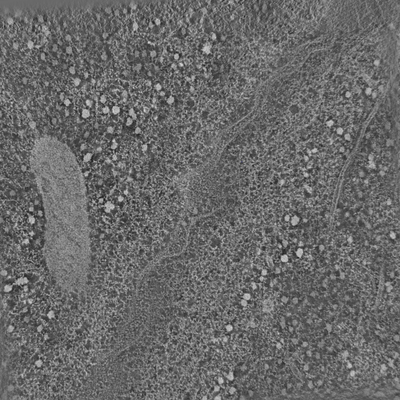 emd_20549.png emd_20549.png | 131.9 KB | ||
| Archive directory |  http://ftp.pdbj.org/pub/emdb/structures/EMD-20549ftp://ftp.pdbj.org/pub/emdb/structures/EMD-20549 http://ftp.pdbj.org/pub/emdb/structures/EMD-20549ftp://ftp.pdbj.org/pub/emdb/structures/EMD-20549 | HTTPS FTP |
-Related structure data

-Links
| EMDB pages | EMDB (EBI/PDBe) / EMDataResource |
|---|
-Map
| File | Download / File: emd_20549.map.gz / Format: CCP4 / Size: 1.4 GB / Type: IMAGE STORED AS SIGNED INTEGER (2 BYTES) | ||||||||||||||||||||||||||||||||||||||||||||||||||||||||||||
|---|---|---|---|---|---|---|---|---|---|---|---|---|---|---|---|---|---|---|---|---|---|---|---|---|---|---|---|---|---|---|---|---|---|---|---|---|---|---|---|---|---|---|---|---|---|---|---|---|---|---|---|---|---|---|---|---|---|---|---|---|---|
| Voxel size | X=Y=Z: 8.1 Å | ||||||||||||||||||||||||||||||||||||||||||||||||||||||||||||
| Density |
| ||||||||||||||||||||||||||||||||||||||||||||||||||||||||||||
| Symmetry | Space group: 1 | ||||||||||||||||||||||||||||||||||||||||||||||||||||||||||||
| Details | EMDB XML:
CCP4 map header:
| ||||||||||||||||||||||||||||||||||||||||||||||||||||||||||||
-Supplemental data
- Sample components
-Entire : Cochlea
| Entire | Name: Cochlea |
|---|---|
| Components |
|
-Supramolecule #1: Cochlea
| Supramolecule | Name: Cochlea / type: tissue / ID: 1 / Parent: 0 |
|---|---|
| Source (natural) | Organism: Cavia porcellus (domestic guinea pig) |
-Experimental details
-Structure determination
| Method | negative staining, cryo EM |
|---|---|
 Processing Processing | electron tomography |
| Aggregation state | tissue |
-Sample preparation
| Buffer | pH: 7 |
|---|---|
| Staining | Type: POSITIVE / Material: Uranyl Acetate |
| Sugar embedding | Material: Epon |
| Grid | Details: unspecified |
| Vitrification | Cryogen name: NITROGEN |
| High pressure freezing | Instrument: OTHER Details: The value given for _emd_high_pressure_freezing.instrument is BALTEC. This is not in a list of allowed values set(['LEICA EM PACT2', 'LEICA EM PACT', 'EMS-002 RAPID IMMERSION FREEZER', ...Details: The value given for _emd_high_pressure_freezing.instrument is BALTEC. This is not in a list of allowed values set(['LEICA EM PACT2', 'LEICA EM PACT', 'EMS-002 RAPID IMMERSION FREEZER', 'OTHER', 'LEICA EM HPM100', 'BAL-TEC HPM 010']) so OTHER is written into the XML file. |
| Sectioning | Ultramicrotomy - Instrument: Leica EM UC6 / Ultramicrotomy - Temperature: 298 K / Ultramicrotomy - Final thickness: 100 nm |
| Fiducial marker | Manufacturer: Electron Microscopy Sciences / Diameter: 5 nm |
- Electron microscopy
| Microscope | FEI/PHILIPS CM200FEG |
|---|---|
| Electron beam | Acceleration voltage: 200 kV / Electron source: FIELD EMISSION GUN |
| Electron optics | Illumination mode: FLOOD BEAM / Imaging mode: BRIGHT FIELDBright-field microscopy |
| Image recording | Film or detector model: TVIPS TEMCAM-F216 (2k x 2k) / Average electron dose: 10.0 e/Å2 |
-Image processing
| Final reconstruction | Number images used: 130 |
|---|
