 ムービー
ムービー コントローラー


+ データを開く

- 基本情報
基本情報
| 登録情報 | データベース: EMDB / ID: EMD-1679 | |||||||||
|---|---|---|---|---|---|---|---|---|---|---|
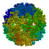
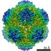
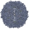
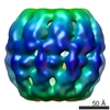
| タイトル | Characterization of the extremophilic, archaeal virus STIV2 | |||||||||
 マップデータ マップデータ | This is a 20 A resolution cryo-EM reconstruction of the archaeal virus STIV2 | |||||||||
 試料 試料 |
| |||||||||
 キーワード キーワード | Archaeal / virus / icosahedral / extremophilic | |||||||||
| 生物種 |   Sulfolobus turreted icosahedral virus 2 (ウイルス) Sulfolobus turreted icosahedral virus 2 (ウイルス) | |||||||||
| 手法 | 単粒子再構成法 / クライオ電子顕微鏡法 / ネガティブ染色法 / 解像度: 20.0 Å | |||||||||
 データ登録者 データ登録者 | Happonen LJ / Redder P / Peng X / Reigstad LJ / Prangishvili D / Butcher SJ | |||||||||
 引用 引用 | ジャーナル: J Virol / 年: 2010 タイトル: Familial relationships in hyperthermo- and acidophilic archaeal viruses. 著者: Lotta Johanna Happonen / Peter Redder / Xu Peng / Laila Johanne Reigstad / David Prangishvili / Sarah Jane Butcher /  要旨: Archaea often live in extreme, harsh environments such as acidic hot springs and hypersaline waters. To date, only two icosahedrally symmetric, membrane-containing archaeal viruses, SH1 and ...Archaea often live in extreme, harsh environments such as acidic hot springs and hypersaline waters. To date, only two icosahedrally symmetric, membrane-containing archaeal viruses, SH1 and Sulfolobus turreted icosahedral virus (STIV), have been described in detail. We report the sequence and three-dimensional structure of a third such virus isolated from a hyperthermoacidophilic crenarchaeon, Sulfolobus strain G4ST-2. Characterization of this new isolate revealed it to be similar to STIV on the levels of genome and structural organization. The genome organization indicates that these two viruses have diverged from a common ancestor. Interestingly, the prominent surface turrets of the two viruses are strikingly different. By sequencing and mass spectrometry, we mapped several large insertions and deletions in the known structural proteins that could account for these differences and showed that both viruses can infect the same host. A combination of genomic and proteomic analyses revealed important new insights into the structural organization of these viruses and added to our limited knowledge of archaeal virus life cycles and host-cell interactions. | |||||||||
| 履歴 |
|
- 構造の表示
構造の表示
| ムービー |
 ムービービューア ムービービューア |
|---|---|
| 構造ビューア | EMマップ: SurfViewMolmilJmol/JSmol |
| 添付画像 |

- ダウンロードとリンク
-EMDBアーカイブ
| マップデータ | emd_1679.map.gz | 50.2 MB | EMDBマップデータ形式 | |
|---|---|---|---|---|
| ヘッダ (付随情報) | emd-1679-v30.xmlemd-1679.xml | 9.2 KB 9.2 KB | 表示 表示 | EMDBヘッダ |
| 画像 | 1679.tif | 739.4 KB | ||
| アーカイブディレクトリ |  http://ftp.pdbj.org/pub/emdb/structures/EMD-1679ftp://ftp.pdbj.org/pub/emdb/structures/EMD-1679 http://ftp.pdbj.org/pub/emdb/structures/EMD-1679ftp://ftp.pdbj.org/pub/emdb/structures/EMD-1679 | HTTPS FTP |
-検証レポート
| 文書・要旨 | emd_1679_validation.pdf.gz | 268.2 KB | 表示 | EMDB検証レポート |
|---|---|---|---|---|
| 文書・詳細版 | emd_1679_full_validation.pdf.gz | 267.3 KB | 表示 | |
| XML形式データ | emd_1679_validation.xml.gz | 7.7 KB | 表示 | |
| アーカイブディレクトリ | https://ftp.pdbj.org/pub/emdb/validation_reports/EMD-1679ftp://ftp.pdbj.org/pub/emdb/validation_reports/EMD-1679 | HTTPS FTP |
-関連構造データ

-リンク
| EMDBのページ | EMDB (EBI/PDBe) / EMDataResource |
|---|
-マップ
| ファイル | ダウンロード / ファイル: emd_1679.map.gz / 形式: CCP4 / 大きさ: 130.3 MB / タイプ: IMAGE STORED AS FLOATING POINT NUMBER (4 BYTES) | ||||||||||||||||||||||||||||||||||||||||||||||||||||||||||||||||||||
|---|---|---|---|---|---|---|---|---|---|---|---|---|---|---|---|---|---|---|---|---|---|---|---|---|---|---|---|---|---|---|---|---|---|---|---|---|---|---|---|---|---|---|---|---|---|---|---|---|---|---|---|---|---|---|---|---|---|---|---|---|---|---|---|---|---|---|---|---|---|
| 注釈 | This is a 20 A resolution cryo-EM reconstruction of the archaeal virus STIV2 | ||||||||||||||||||||||||||||||||||||||||||||||||||||||||||||||||||||
| 投影像・断面図 | 画像のコントロール
画像は Spider により作成 | ||||||||||||||||||||||||||||||||||||||||||||||||||||||||||||||||||||
| ボクセルのサイズ | X=Y=Z: 4.42 Å | ||||||||||||||||||||||||||||||||||||||||||||||||||||||||||||||||||||
| 密度 |
| ||||||||||||||||||||||||||||||||||||||||||||||||||||||||||||||||||||
| 対称性 | 空間群: 1 | ||||||||||||||||||||||||||||||||||||||||||||||||||||||||||||||||||||
| 詳細 | EMDB XML:
CCP4マップ ヘッダ情報:
| ||||||||||||||||||||||||||||||||||||||||||||||||||||||||||||||||||||
 Z (Sec.)
 Y (Row.)
 X (Col.)


-添付データ
- 試料の構成要素
-全体 : STIV2 virus
| 全体 | 名称: STIV2 virus |
|---|---|
| 要素 |
|
-超分子 #1000: STIV2 virus
| 超分子 | 名称: STIV2 virus / タイプ: sample / ID: 1000 / Number unique components: 1 |
|---|
-超分子 #1: Sulfolobus turreted icosahedral virus 2
| 超分子 | 名称: Sulfolobus turreted icosahedral virus 2 / タイプ: virus / ID: 1 / Name.synonym: STIV2 / NCBI-ID: 754004 / 生物種: Sulfolobus turreted icosahedral virus 2 / ウイルスタイプ: VIRION / ウイルス・単離状態: STRAIN / ウイルス・エンベロープ: No / ウイルス・中空状態: No / Syn species name: STIV2 |
|---|---|
| 宿主 | 生物種:  Sulfolobus islandicus (古細菌) / 別称: ARCHAEA Sulfolobus islandicus (古細菌) / 別称: ARCHAEA |
| ウイルス殻 | Shell ID: 1 / 直径: 710 Å / T番号(三角分割数): 31 |
-実験情報
-構造解析
| 手法 | ネガティブ染色法, クライオ電子顕微鏡法 |
|---|---|
 解析 解析 | 単粒子再構成法 |
| 試料の集合状態 | particle |
-試料調製
| 緩衝液 | pH: 3.5 / 詳細: 50 mM sodium citrate, pH 3.5 |
|---|---|
| 染色 | タイプ: NEGATIVE 詳細: Vitrified. Grids were blotted for roughly one second before being plunged into liquid ethane. |
| グリッド | 詳細: Quantifoil-grids |
| 凍結 | 凍結剤: ETHANE / 装置: HOMEMADE PLUNGER / 詳細: Vitrification instrument: Guillotine 手法: A small vial of ethane is placed inside a larger liquid nitrogen reservoir. The grid holding 3 microliters of the sample is held in place at the bottom of a plunger by the means of fine ...手法: A small vial of ethane is placed inside a larger liquid nitrogen reservoir. The grid holding 3 microliters of the sample is held in place at the bottom of a plunger by the means of fine tweezers. When the liquid ethane is ready, a piece of filter paper is then pressed against the sample to blot off excess buffer, sufficient to leave a thin layer on the grid. The filter paper is removed, and the plunger is allowed to drop into the liquid ethane. Once the grid enters the liquid ethane, the sample is rapidly frozen, and the grid is transferred under liquid nitrogen to a storage box immersed in liquid nitrogen for later use in the microscope. |
- 電子顕微鏡法
| 顕微鏡 | FEI TECNAI F20 |
|---|---|
| 詳細 | Low dose conditions |
| 撮影 | カテゴリ: CCD フィルム・検出器のモデル: GATAN ULTRASCAN 4000 (4k x 4k) デジタル化 - サンプリング間隔: 4.42 µm / 実像数: 358 / 平均電子線量: 18 e/Å2 |
| 電子線 | 加速電圧: 200 kV / 電子線源:  FIELD EMISSION GUN FIELD EMISSION GUN |
| 電子光学系 | 倍率(補正後): 66400 / 照射モード: FLOOD BEAM / 撮影モード: BRIGHT FIELD / Cs: 2.0 mm / 最大 デフォーカス(公称値): 5.2 µm / 最小 デフォーカス(公称値): 0.5 µm / 倍率(公称値): 68000 |
| 試料ステージ | 試料ホルダー: Side entry liquid nitrogen-cooled side entry holder 試料ホルダーモデル: GATAN LIQUID NITROGEN |
| 実験機器 |  モデル: Tecnai F20 / 画像提供: FEI Company |
-画像解析
| CTF補正 | 詳細: Each micrograph |
|---|---|
| 最終 再構成 | 想定した対称性 - 点群: I (正20面体型対称) / アルゴリズム: OTHER / 解像度のタイプ: BY AUTHOR / 解像度: 20.0 Å / 解像度の算出法: FSC 0.5 CUT-OFF / ソフトウェア - 名称: PFT, POR, EM3DR2, P3DR / 使用した粒子像数: 713 |
