National Institutes of Health/National Institute Of Allergy and Infectious Diseases (NIH/NIAID)
R01 AI056346
United States
National Natural Science Foundation of China (NSFC)
313290002
China
National Institutes of Health/National Center for Research Resources (NIH/NCRR)
1S10RR23057
United States
China Academy of Sciences
XDB02050000
China
National Science Foundation (NSF, United States)
DBI-1338135
United States
Citation
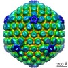
Journal: J Gen Virol / Year: 2017 Title: A pUL25 dimer interfaces the pseudorabies virus capsid and tegument. Authors: Yun-Tao Liu / Jiansen Jiang / Kevin Patrick Bohannon / Xinghong Dai / G W Gant Luxton / Wong Hoi Hui / Guo-Qiang Bi / Gregory Allan Smith / Z Hong Zhou / Abstract: Inside the virions of α-herpesviruses, tegument protein pUL25 anchors the tegument to capsid vertices through direct interactions with tegument proteins pUL17 and pUL36. In addition to promoting ...Inside the virions of α-herpesviruses, tegument protein pUL25 anchors the tegument to capsid vertices through direct interactions with tegument proteins pUL17 and pUL36. In addition to promoting virion assembly, both pUL25 and pUL36 are critical for intracellular microtubule-dependent capsid transport. Despite these essential roles during infection, the stoichiometry and precise organization of pUL25 and pUL36 on the capsid surface remain controversial due to the insufficient resolution of existing reconstructions from cryo-electron microscopy (cryoEM). Here, we report a three-dimensional (3D) icosahedral reconstruction of pseudorabies virus (PRV), a varicellovirus of the α-herpesvirinae subfamily, obtained by electron-counting cryoEM at 4.9 Å resolution. Our reconstruction resolves a dimer of pUL25 forming a capsid-associated tegument complex with pUL36 and pUL17 through a coiled coil helix bundle, thus correcting previous misinterpretations. A comparison between reconstructions of PRV and the γ-herpesvirus Kaposi's sarcoma-associated herpesvirus (KSHV) reinforces their similar architectures and establishes important subfamily differences in the capsid-tegument interface.
History
Deposition
Jun 8, 2017
-
Header (metadata) release
Jul 19, 2017
-
Map release
Nov 1, 2017
-
Update
Dec 25, 2019
-
Current status
Dec 25, 2019
Processing site: RCSB / Status: Released
-
Structure visualization
Movie
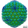
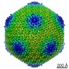
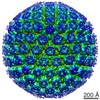
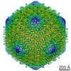
Surface view with section colored by density value
Film or detector model: GATAN K2 SUMMIT (4k x 4k) / Detector mode: SUPER-RESOLUTION / Digitization - Frames/image: 2-25 / Number real images: 1830 / Average electron dose: 18.0 e/Å2
Experimental equipment
Model: Titan Krios / Image courtesy: FEI Company
-
Image processing
Particle selection
Number selected: 13537
CTF correction
Software: (Name: CTFFIND (ver. 3), RELION)
Startup model
Type of model: INSILICO MODEL In silico model: The particles were processed using EMAN for initial 3D reconstruction and refinement with icosahedral symmetry enforced.
Initial angle assignment
Type: NOT APPLICABLE
Final angle assignment
Type: NOT APPLICABLE
Final reconstruction
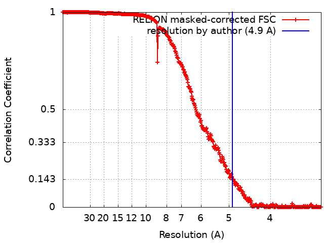
Applied symmetry - Point group: I (icosahedral) / Resolution.type: BY AUTHOR / Resolution: 4.9 Å / Resolution method: FSC 0.143 CUT-OFF / Software - Name: RELION / Number images used: 13537
FSC plot (resolution estimation)
-
Atomic model buiding 1
Refinement
Protocol: RIGID BODY FIT
+
About Yorodumi
-
News
-
Feb 9, 2022. New format data for meta-information of EMDB entries
New format data for meta-information of EMDB entries
Version 3 of the EMDB header file is now the official format.
The previous official version 1.9 will be removed from the archive.
In the structure databanks used in Yorodumi, some data are registered as the other names, "COVID-19 virus" and "2019-nCoV". Here are the details of the virus and the list of structure data.
Jan 31, 2019. EMDB accession codes are about to change! (news from PDBe EMDB page)
EMDB accession codes are about to change! (news from PDBe EMDB page)
The allocation of 4 digits for EMDB accession codes will soon come to an end. Whilst these codes will remain in use, new EMDB accession codes will include an additional digit and will expand incrementally as the available range of codes is exhausted. The current 4-digit format prefixed with “EMD-” (i.e. EMD-XXXX) will advance to a 5-digit format (i.e. EMD-XXXXX), and so on. It is currently estimated that the 4-digit codes will be depleted around Spring 2019, at which point the 5-digit format will come into force.
The EM Navigator/Yorodumi systems omit the EMD- prefix.
Related info.:Q: What is EMD? / ID/Accession-code notation in Yorodumi/EM Navigator
Yorodumi is a browser for structure data from EMDB, PDB, SASBDB, etc.
This page is also the successor to EM Navigator detail page, and also detail information page/front-end page for Omokage search.
The word "yorodu" (or yorozu) is an old Japanese word meaning "ten thousand". "mi" (miru) is to see.
Related info.:EMDB / PDB / SASBDB / Comparison of 3 databanks / Yorodumi Search / Aug 31, 2016. New EM Navigator & Yorodumi / Yorodumi Papers / Jmol/JSmol / Function and homology information / Changes in new EM Navigator and Yorodumi
 Movie
Movie Controller


 Open data
 Basic information
 Map data
 Sample
 Suid herpesvirus 1 (strain Becker)
 single particle reconstruction /
  Authors
 United States,
  China, 5 items
  Citation
 Structure visualization
 Movie viewer
 Downloads & links
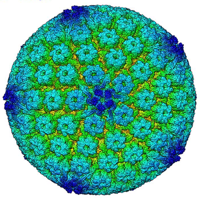 emd_8760.png
 http://ftp.pdbj.org/pub/emdb/structures/EMD-8760


 Sample components
 Processing
 Electron microscopy