 Movie
Movie Controller

[English] 日本語
 Yorodumi
- EMDB-8730: Cryo-EM reconstruction of the HIV-1 BG505 SOSIP.664 Env trimer in... -
+ Open data

- Basic information
| Entry | Database: EMDB / ID: EMD-8730 | ||||||||||||
|---|---|---|---|---|---|---|---|---|---|---|---|---|---|
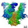
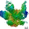
| Title | Cryo-EM reconstruction of the HIV-1 BG505 SOSIP.664 Env trimer in complex with soluble CD4 (D1-D2) and fragment antigen binding variable domain of 17b | ||||||||||||
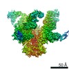
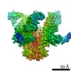
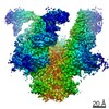
 Map data Map data | Complex formed by HIV-1 BG505 SOSIP.664 Env with soluble CD4 (2-domain) and broadly neutralizing antibody 17b | ||||||||||||
 Sample Sample |
| ||||||||||||
| Function / homology |  Function and homology information Function and homology informationpositive regulation of plasma membrane raft polarization / positive regulation of receptor clustering / positive regulation of establishment of T cell polarity / virus-mediated perturbation of host defense response / host cell endosome membrane / clathrin-dependent endocytosis of virus by host cell /  viral protein processing / fusion of virus membrane with host plasma membrane / fusion of virus membrane with host endosome membrane / viral envelope ...positive regulation of plasma membrane raft polarization / positive regulation of receptor clustering / positive regulation of establishment of T cell polarity / virus-mediated perturbation of host defense response / host cell endosome membrane / clathrin-dependent endocytosis of virus by host cell / viral protein processing / fusion of virus membrane with host plasma membrane / fusion of virus membrane with host endosome membrane / viral envelope / structural molecule activity / virion attachment to host cell / host cell plasma membrane / virion membrane / plasma membrane viral protein processing / fusion of virus membrane with host plasma membrane / fusion of virus membrane with host endosome membrane / viral envelope ...positive regulation of plasma membrane raft polarization / positive regulation of receptor clustering / positive regulation of establishment of T cell polarity / virus-mediated perturbation of host defense response / host cell endosome membrane / clathrin-dependent endocytosis of virus by host cell / viral protein processing / fusion of virus membrane with host plasma membrane / fusion of virus membrane with host endosome membrane / viral envelope / structural molecule activity / virion attachment to host cell / host cell plasma membrane / virion membrane / plasma membraneSimilarity search - Function | ||||||||||||
| Biological species |   Human immunodeficiency virus 1 / Human immunodeficiency virus 1 /  Homo sapiens (human) Homo sapiens (human) | ||||||||||||
| Method | single particle reconstruction / cryo EM / Resolution: 8.6 Å | ||||||||||||
 Authors Authors | Lyumkis D / de Val N / Ward AB | ||||||||||||
| Funding support |  United States, 3 items United States, 3 items
| ||||||||||||
 Citation Citation | Journal: Nature / Year: 2017 Title: Open and closed structures reveal allostery and pliability in the HIV-1 envelope spike. Authors: Gabriel Ozorowski / Jesper Pallesen / Natalia de Val / Dmitry Lyumkis / Christopher A Cottrell / Jonathan L Torres / Jeffrey Copps / Robyn L Stanfield / Albert Cupo / Pavel Pugach / John P ...Authors: Gabriel Ozorowski / Jesper Pallesen / Natalia de Val / Dmitry Lyumkis / Christopher A Cottrell / Jonathan L Torres / Jeffrey Copps / Robyn L Stanfield / Albert Cupo / Pavel Pugach / John P Moore / Ian A Wilson / Andrew B Ward / Abstract: For many enveloped viruses, binding to a receptor(s) on a host cell acts as the first step in a series of events culminating in fusion with the host cell membrane and transfer of genetic material for ...For many enveloped viruses, binding to a receptor(s) on a host cell acts as the first step in a series of events culminating in fusion with the host cell membrane and transfer of genetic material for replication. The envelope glycoprotein (Env) trimer on the surface of HIV is responsible for receptor binding and fusion. Although Env can tolerate a high degree of mutation in five variable regions (V1-V5), and also at N-linked glycosylation sites that contribute roughly half the mass of Env, the functional sites for recognition of receptor CD4 and co-receptor CXCR4/CCR5 are conserved and essential for viral fitness. Soluble SOSIP Env trimers are structural and antigenic mimics of the pre-fusion native, surface-presented Env, and are targets of broadly neutralizing antibodies. Thus, they are attractive immunogens for vaccine development. Here we present high-resolution cryo-electron microscopy structures of subtype B B41 SOSIP Env trimers in complex with CD4 and antibody 17b, or with antibody b12, at resolutions of 3.7 Å and 3.6 Å, respectively. We compare these to cryo-electron microscopy reconstructions of B41 SOSIP Env trimers with no ligand or in complex with either CD4 or the CD4-binding-site antibody PGV04 at 5.6 Å, 5.2 Å and 7.4 Å resolution, respectively. Consequently, we present the most complete description yet, to our knowledge, of the CD4-17b-induced intermediate and provide the molecular basis of the receptor-binding-induced conformational change required for HIV-1 entry into host cells. Both CD4 and b12 induce large, previously uncharacterized conformational rearrangements in the gp41 subunits, and the fusion peptide becomes buried in a newly formed pocket. These structures provide key details on the biological function of the type I viral fusion machine from HIV-1 as well as new templates for inhibitor design. | ||||||||||||
| History |
|
- Structure visualization
| Movie |
Movie viewer |
|---|---|
| Structure viewer | EM map: SurfViewMolmilJmol/JSmol |
| Supplemental images |

- Downloads & links
-EMDB archive
| Map data | emd_8730.map.gz | 6.6 MB | EMDB map data format | |
|---|---|---|---|---|
| Header (meta data) | emd-8730-v30.xmlemd-8730.xml | 24 KB 24 KB | Display Display | EMDB header |
| Images | 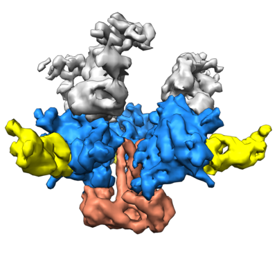 emd_8730.png emd_8730.png | 129.9 KB | ||
| Archive directory |  http://ftp.pdbj.org/pub/emdb/structures/EMD-8730ftp://ftp.pdbj.org/pub/emdb/structures/EMD-8730 http://ftp.pdbj.org/pub/emdb/structures/EMD-8730ftp://ftp.pdbj.org/pub/emdb/structures/EMD-8730 | HTTPS FTP |
-Related structure data
| Related structure data |  8713C  8714C  8715C 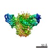 8716C 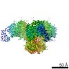 8717C 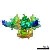 8729C 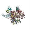 5vn3C 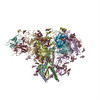 5vn8C C: citing same article ( |
|---|---|
| Similar structure data |

-Links
| EMDB pages | EMDB (EBI/PDBe) / EMDataResource |
|---|---|
| Related items in Molecule of the Month |


-Map
| File | Download / File: emd_8730.map.gz / Format: CCP4 / Size: 8 MB / Type: IMAGE STORED AS FLOATING POINT NUMBER (4 BYTES) | ||||||||||||||||||||||||||||||||||||||||||||||||||||||||||||||||||||
|---|---|---|---|---|---|---|---|---|---|---|---|---|---|---|---|---|---|---|---|---|---|---|---|---|---|---|---|---|---|---|---|---|---|---|---|---|---|---|---|---|---|---|---|---|---|---|---|---|---|---|---|---|---|---|---|---|---|---|---|---|---|---|---|---|---|---|---|---|---|
| Annotation | Complex formed by HIV-1 BG505 SOSIP.664 Env with soluble CD4 (2-domain) and broadly neutralizing antibody 17b | ||||||||||||||||||||||||||||||||||||||||||||||||||||||||||||||||||||
| Voxel size | X=Y=Z: 2.62 Å | ||||||||||||||||||||||||||||||||||||||||||||||||||||||||||||||||||||
| Density |
| ||||||||||||||||||||||||||||||||||||||||||||||||||||||||||||||||||||
| Symmetry | Space group: 1 | ||||||||||||||||||||||||||||||||||||||||||||||||||||||||||||||||||||
| Details | EMDB XML:
CCP4 map header:
| ||||||||||||||||||||||||||||||||||||||||||||||||||||||||||||||||||||
-Supplemental data
- Sample components
-Entire : HIV-1 Env BG505 SOSIP.664 in complex with soluble CD4 (2-domain) ...
| Entire | Name: HIV-1 Env BG505 SOSIP.664 in complex with soluble CD4 (2-domain) and Fab domain of neutralizing 17b antibody |
|---|---|
| Components |
|
-Supramolecule #1: HIV-1 Env BG505 SOSIP.664 in complex with soluble CD4 (2-domain) ...
| Supramolecule | Name: HIV-1 Env BG505 SOSIP.664 in complex with soluble CD4 (2-domain) and Fab domain of neutralizing 17b antibody type: complex / ID: 1 / Parent: 0 / Macromolecule list: all |
|---|---|
| Source (natural) | Organism: Human immunodeficiency virus 1 |
| Molecular weight | Theoretical: 150 KDa |
-Supramolecule #2: HIV-1 Env BG505 SOSIP.664
| Supramolecule | Name: HIV-1 Env BG505 SOSIP.664 / type: complex / ID: 2 / Parent: 1 / Macromolecule list: #1-#2 |
|---|---|
| Source (natural) | Organism: Human immunodeficiency virus 1 |
| Recombinant expression | Organism: Homo sapiens (human) / Recombinant cell: HEK293F |
-Supramolecule #3: CD4 (2-domain)
| Supramolecule | Name: CD4 (2-domain) / type: complex / ID: 3 / Parent: 1 / Macromolecule list: #3 |
|---|---|
| Source (natural) | Organism: Homo sapiens (human) |
| Recombinant expression | Organism: Homo sapiens (human) / Recombinant cell: HEK293F |
-Supramolecule #4: broadly neutralizing antibody (Fab) 17b
| Supramolecule | Name: broadly neutralizing antibody (Fab) 17b / type: complex / ID: 4 / Parent: 1 / Macromolecule list: #4-#5 |
|---|---|
| Source (natural) | Organism: Homo sapiens (human) |
| Recombinant expression | Organism: Homo sapiens (human) / Recombinant cell: HEK293F |
-Macromolecule #1: HIV-1 Env BG505 SOSIP.664 gp41
| Macromolecule | Name: HIV-1 Env BG505 SOSIP.664 gp41 / type: protein_or_peptide / ID: 1 / Enantiomer: LEVO |
|---|---|
| Source (natural) | Organism: Human immunodeficiency virus 1 |
| Recombinant expression | Organism: Homo sapiens (human) |
| Sequence | String: AVGIGAVFLG FLGAAGSTMG AASMTLTVQA RNLLSGIVQQ QSNLLRAPEA QQHLLKLTVW GIKQLQARVL AVERYLRDQQ LLGIWGCSGK LICCTNVPWN SSWSNRNLSE IWDNMTWLQW DKEISNYTQI IYGLLEESQN QQEKNEQDLL ALD |
-Macromolecule #2: HIV-1 Env BG505 SOSIP.664 gp120
| Macromolecule | Name: HIV-1 Env BG505 SOSIP.664 gp120 / type: protein_or_peptide / ID: 2 / Enantiomer: LEVO |
|---|---|
| Source (natural) | Organism: Human immunodeficiency virus 1 |
| Recombinant expression | Organism: Homo sapiens (human) |
| Sequence | String: MDAMKRGLCC VLLLCGAVFV SPSQEIHARF RRGARAENLW VTVYYGVPVW KDAETTLFCA SDAKAYETEK HNVWATHACV PTDPNPQEIH LENVTEEFNM WKNNMVEQMH TDIISLWDQS LKPCVKLTPL CVTLQCTNVT NNITDDMRGE LKNCSFNMTT ELRDKKQKVY ...String: MDAMKRGLCC VLLLCGAVFV SPSQEIHARF RRGARAENLW VTVYYGVPVW KDAETTLFCA SDAKAYETEK HNVWATHACV PTDPNPQEIH LENVTEEFNM WKNNMVEQMH TDIISLWDQS LKPCVKLTPL CVTLQCTNVT NNITDDMRGE LKNCSFNMTT ELRDKKQKVY SLFYRLDVVQ INENQGNRSN NSNKEYRLIN CNTSAITQAC PKVSFEPIPI HYCAPAGFAI LKCKDKKFNG TGPCPSVSTV QCTHGIKPVV STQLLLNGSL AEEEVMIRSE NITNNAKNIL VQFNTPVQIN CTRPNNNTRK SIRIGPGQAF YATGDIIGDI RQAHCNVSKA TWNETLGKVV KQLRKHFGNN TIIRFANSSG GDLEVTTHSF NCGGEFFYCN TSGLFNSTWI SNTSVQGSNS TGSNDSITLP CRIKQIINMW QRIGQAMYAP PIQGVIRCVS NITGLILTRD GGSTNSTTET FRPGGGDMRD NWRSELYKYK VVKIEPLGVA PTRCKRRVVG RRRRRR |
-Macromolecule #3: soluble CD4 (2-domain)
| Macromolecule | Name: soluble CD4 (2-domain) / type: protein_or_peptide / ID: 3 / Enantiomer: LEVO |
|---|---|
| Source (natural) | Organism: Homo sapiens (human) |
| Recombinant expression | Organism: Homo sapiens (human) |
| Sequence | String: KKVVLGKKGD TVELTCTASQ KKSIQFHWKN SNQIKILGNQ GSFLTKGPSK LNDRADSRRS LWDQGNFPLI IKNLKIEDSD TYICEVEDQK EEVQLLVFGL TANSDTHLLQ GQSLTLTLES PPGSSPSVQC RSPRGKNIQG GKTLSVSQLE LQDSGTWTCT VLQNQKKVEF KIDIVV |
-Macromolecule #4: broadly neutralizing antibody (Fab) 17b light chain
| Macromolecule | Name: broadly neutralizing antibody (Fab) 17b light chain / type: protein_or_peptide / ID: 4 / Enantiomer: LEVO |
|---|---|
| Source (natural) | Organism: Homo sapiens (human) |
| Recombinant expression | Organism: Homo sapiens (human) |
| Sequence | String: ELELTQSPAT LSVSPGERAT LSCRASESVS SDLAWYQQKP GQAPRLLIYG ASTRATGVPA RFSGSGSGAE FTLTISSLQS EDFAVYYCQQ YNNWPPRYTF GQGTRLEIKR |
-Macromolecule #5: broadly neutralizing antibody (Fab) 17b heavy chain
| Macromolecule | Name: broadly neutralizing antibody (Fab) 17b heavy chain / type: protein_or_peptide / ID: 5 / Enantiomer: LEVO |
|---|---|
| Source (natural) | Organism: Homo sapiens (human) |
| Recombinant expression | Organism: Homo sapiens (human) |
| Sequence | String: QVQLLESGAE VKKPGSSVKV SCKASGDTFI RYSFTWVRQA PGQGLEWMGR IITILDVAHY APHLQGRVTI TADKSTSTVY LELRNLRSDD TAVYFCAGVY EGEADEGEYD NNGFLKHWGQ GTLVTVT |
-Experimental details
-Structure determination
| Method | cryo EM |
|---|---|
 Processing Processing | single particle reconstruction |
| Aggregation state | particle |
-Sample preparation
| Concentration | 1 mg/mL | ||||||||||||
|---|---|---|---|---|---|---|---|---|---|---|---|---|---|
| Buffer | pH: 7.4 Component:
| ||||||||||||
| Grid | Model: C-flat / Material: COPPER / Mesh: 400 / Support film - Material: CARBON / Support film - topology: HOLEY / Pretreatment - Type: PLASMA CLEANING / Pretreatment - Atmosphere: OTHER | ||||||||||||
| Vitrification | Cryogen name: ETHANE / Chamber humidity: 50 % / Chamber temperature: 277 K / Instrument: HOMEMADE PLUNGER Details: 5 microliters of the complex was incubated with 3 microliters of a fresh 1.8 mM DDM solution. A 3 microliter aliquot of the complex was applied to a C-Flat grid (CF-2/2-4C, Electron ...Details: 5 microliters of the complex was incubated with 3 microliters of a fresh 1.8 mM DDM solution. A 3 microliter aliquot of the complex was applied to a C-Flat grid (CF-2/2-4C, Electron Microscopy Sciences, Protochips, Inc.) which had been plasma cleaned for 5 seconds using a mixture of Ar/O2 (Gatan Solarus 950 Plasma system), blotted off, and then immediately plunged into liquid ethane using a manual freeze plunger.. |
- Electron microscopy
| Microscope | FEI TITAN KRIOS |
|---|---|
| Electron beam | Acceleration voltage: 300 kV / Electron source: FIELD EMISSION GUN |
| Electron optics | C2 aperture diameter: 100.0 µm / Calibrated defocus max: 4.0 µm / Calibrated defocus min: 1.0 µm / Calibrated magnification: 38167 / Illumination mode: FLOOD BEAM / Imaging mode: BRIGHT FIELDBright-field microscopy / Cs: 2.7 mm / Nominal magnification: 22500 |
| Sample stage | Specimen holder model: FEI TITAN KRIOS AUTOGRID HOLDER / Cooling holder cryogen: NITROGEN |
| Temperature | Min: 90.0 K / Max: 90.0 K |
| Image recording | Film or detector model: GATAN K2 SUMMIT (4k x 4k) / Detector mode: COUNTING / Digitization - Dimensions - Width: 3838 pixel / Digitization - Dimensions - Height: 3710 pixel / Digitization - Sampling interval: 5.0 µm / Digitization - Frames/image: 1-28 / Number grids imaged: 1 / Average exposure time: 5.6 sec. / Average electron dose: 32.0 e/Å2 Details: Individual frames were gain-corrected, aligned, and summed using MotionCor. |
| Experimental equipment |  Model: Titan Krios / Image courtesy: FEI Company |
-Image processing
| CTF correction | Software - Name: CTFFIND (ver. 3) / Details: performed internally in Relion and Frealign |
|---|---|
| Startup model | Type of model: INSILICO MODEL / In silico model: common lines model using OptiMod Details: An initial model was generated directly from the class averages using OptiMod. |
| Initial angle assignment | Type: PROJECTION MATCHING Projection matching processing - Angular sampling: 7.5 degrees Software - Name: RELION (ver. 1.3) / Details: Relion 3D classification, auto mode |
| Final 3D classification | Number classes: 4 / Software - Name: FREALIGN (ver. 9.11) |
| Final angle assignment | Type: PROJECTION MATCHING / Software - Name: FREALIGN (ver. 9.11) / Details: Frealign 3D classification and refinement |
| Final reconstruction | Applied symmetry - Point group: C3 (3 fold cyclic) / Algorithm: FOURIER SPACE / Resolution.type: BY AUTHOR / Resolution: 8.6 Å / Resolution method: FSC 0.143 CUT-OFF / Software - Name: FREALIGN (ver. 9.11) / Details: Resolution-limited refinement used throughout / Number images used: 5716 |
