glutamate-gated calcium ion channel activity / NMDA glutamate receptor activity / NMDA selective glutamate receptor complex / calcium ion transmembrane import into cytosol / protein heterotetramerization / response to zinc ion / response to magnesium ion / late endosome / postsynaptic membrane / lysosome ...glutamate-gated calcium ion channel activity / NMDA glutamate receptor activity / NMDA selective glutamate receptor complex / calcium ion transmembrane import into cytosol / protein heterotetramerization / response to zinc ion / response to magnesium ion / late endosome / postsynaptic membrane / lysosome / metal ion binding / plasma membrane Similarity search - Function
Glutamate [NMDA] receptor, epsilon subunit, C-terminal / N-methyl D-aspartate receptor 2B3 C-terminus / Bacterial extracellular solute-binding proteins, family 3 / Solute-binding protein family 3/N-terminal domain of MltF / Ionotropic glutamate receptor, metazoa / Ligated ion channel L-glutamate- and glycine-binding site / : / Ligand-gated ion channel / Ionotropic glutamate receptor, L-glutamate and glycine-binding domain / Ligated ion channel L-glutamate- and glycine-binding site ...Glutamate [NMDA] receptor, epsilon subunit, C-terminal / N-methyl D-aspartate receptor 2B3 C-terminus / Bacterial extracellular solute-binding proteins, family 3 / Solute-binding protein family 3/N-terminal domain of MltF / Ionotropic glutamate receptor, metazoa / Ligated ion channel L-glutamate- and glycine-binding site / : / Ligand-gated ion channel / Ionotropic glutamate receptor, L-glutamate and glycine-binding domain / Ligated ion channel L-glutamate- and glycine-binding site / Ionotropic glutamate receptor / Eukaryotic homologues of bacterial periplasmic substrate binding proteins. / Receptor, ligand binding region / Receptor family ligand binding region / Periplasmic binding protein-like I Similarity search - Domain/homology
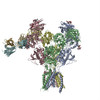
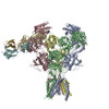
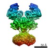
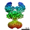
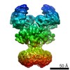
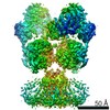
Journal: Science / Year: 2017 Title: Cryo-EM structures of the triheteromeric NMDA receptor and its allosteric modulation. Authors: Wei Lü / Juan Du / April Goehring / Eric Gouaux / Abstract: -methyl-d-aspartate receptors (NMDARs) are heterotetrameric ion channels assembled as diheteromeric or triheteromeric complexes. Here, we report structures of the triheteromeric GluN1/GluN2A/GluN2B ...-methyl-d-aspartate receptors (NMDARs) are heterotetrameric ion channels assembled as diheteromeric or triheteromeric complexes. Here, we report structures of the triheteromeric GluN1/GluN2A/GluN2B receptor in the absence or presence of the GluN2B-specific allosteric modulator Ro 25-6981 (Ro), determined by cryogenic electron microscopy (cryo-EM). In the absence of Ro, the GluN2A and GluN2B amino-terminal domains (ATDs) adopt "closed" and "open" clefts, respectively. Upon binding Ro, the GluN2B ATD clamshell transitions from an open to a closed conformation. Consistent with a predominance of the GluN2A subunit in ion channel gating, the GluN2A subunit interacts more extensively with GluN1 subunits throughout the receptor, in comparison with the GluN2B subunit. Differences in the conformation of the pseudo-2-fold-related GluN1 subunits further reflect receptor asymmetry. The triheteromeric NMDAR structures provide the first view of the most common NMDA receptor assembly and show how incorporation of two different GluN2 subunits modifies receptor symmetry and subunit interactions, allowing each subunit to uniquely influence receptor structure and function, thus increasing receptor complexity.
History
Deposition
Feb 1, 2017
-
Header (metadata) release
Feb 22, 2017
-
Map release
Mar 22, 2017
-
Update
Apr 11, 2018
-
Current status
Apr 11, 2018
Processing site: RCSB / Status: Released
-
Structure visualization
Movie
Surface view with section colored by density value
In the structure databanks used in Yorodumi, some data are registered as the other names, "COVID-19 virus" and "2019-nCoV". Here are the details of the virus and the list of structure data.
Jan 31, 2019. EMDB accession codes are about to change! (news from PDBe EMDB page)
EMDB accession codes are about to change! (news from PDBe EMDB page)
The allocation of 4 digits for EMDB accession codes will soon come to an end. Whilst these codes will remain in use, new EMDB accession codes will include an additional digit and will expand incrementally as the available range of codes is exhausted. The current 4-digit format prefixed with “EMD-” (i.e. EMD-XXXX) will advance to a 5-digit format (i.e. EMD-XXXXX), and so on. It is currently estimated that the 4-digit codes will be depleted around Spring 2019, at which point the 5-digit format will come into force.
The EM Navigator/Yorodumi systems omit the EMD- prefix.
Related info.:Q: What is EMD? / ID/Accession-code notation in Yorodumi/EM Navigator
Yorodumi is a browser for structure data from EMDB, PDB, SASBDB, etc.
This page is also the successor to EM Navigator detail page, and also detail information page/front-end page for Omokage search.
The word "yorodu" (or yorozu) is an old Japanese word meaning "ten thousand". "mi" (miru) is to see.
Related info.:EMDB / PDB / SASBDB / Comparison of 3 databanks / Yorodumi Search / Aug 31, 2016. New EM Navigator & Yorodumi / Yorodumi Papers / Jmol/JSmol / Function and homology information / Changes in new EM Navigator and Yorodumi
 Movie
Movie Controller


 Open data
 Basic information
 Map data
 Sample
 Function and homology information
 Authors
 Citation
 Structure visualization
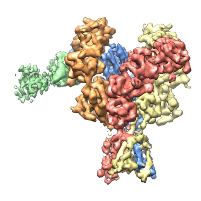
 Downloads & links
 emd_8580.png
 http://ftp.pdbj.org/pub/emdb/structures/EMD-8580


 Z (Sec.)
 Y (Row.)
 X (Col.)


 Sample components
 Processing
 Electron microscopy
