 Movie
Movie Controller


+ Open data

- Basic information
| Entry | Database: EMDB / ID: EMD-3878 | |||||||||
|---|---|---|---|---|---|---|---|---|---|---|
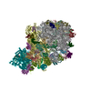
| Title | The distal end of a non-contractile T6SS sheath. | |||||||||
 Map data Map data | The distal end of a non-contractile T6SS sheath. | |||||||||
 Sample Sample |
| |||||||||
| Biological species |    Vibrio cholerae (bacteria) Vibrio cholerae (bacteria) | |||||||||
| Method | single particle reconstruction / cryo EM / Resolution: 7.5 Å | |||||||||
 Authors Authors | Nazarov S / Basler M | |||||||||
| Funding support |  Switzerland, 1 items Switzerland, 1 items
| |||||||||
 Citation Citation | Journal: EMBO J / Year: 2018 Title: Cryo-EM reconstruction of Type VI secretion system baseplate and sheath distal end. Authors: Sergey Nazarov / Johannes P Schneider / Maximilian Brackmann / Kenneth N Goldie / Henning Stahlberg / Marek Basler / Abstract: The bacterial Type VI secretion system (T6SS) assembles from three major parts: a membrane complex that spans inner and outer membranes, a baseplate, and a sheath-tube polymer. The baseplate ...The bacterial Type VI secretion system (T6SS) assembles from three major parts: a membrane complex that spans inner and outer membranes, a baseplate, and a sheath-tube polymer. The baseplate assembles around a tip complex with associated effectors and connects to the membrane complex by TssK. The baseplate assembly initiates sheath-tube polymerization, which in some organisms requires TssA. Here, we analyzed both ends of isolated non-contractile sheaths by cryo-electron microscopy. Our analysis suggests that the baseplate, solved to an average 8.0 Å resolution, is composed of six subunits of TssE/F/G and the baseplate periphery is decorated by six TssK trimers. The VgrG/PAAR tip complex in the center of the baseplate is surrounded by a cavity, which may accommodate up to ~450 kDa of effector proteins. The distal end of the sheath, resolved to an average 7.5 Å resolution, shows sixfold symmetry; however, its protein composition is unclear. Our structures provide an important step toward an atomic model of the complete T6SS assembly. | |||||||||
| History |
|
- Structure visualization
| Movie |
 Movie viewer Movie viewer |
|---|---|
| Structure viewer | EM map: SurfViewMolmilJmol/JSmol |
| Supplemental images |

- Downloads & links
-EMDB archive
| Map data | emd_3878.map.gz | 48.5 MB | EMDB map data format | |
|---|---|---|---|---|
| Header (meta data) | emd-3878-v30.xmlemd-3878.xml | 17.4 KB 17.4 KB | Display Display | EMDB header |
| FSC (resolution estimation) | emd_3878_fsc.xml | 9.2 KB | Display | FSC data file |
| Images | 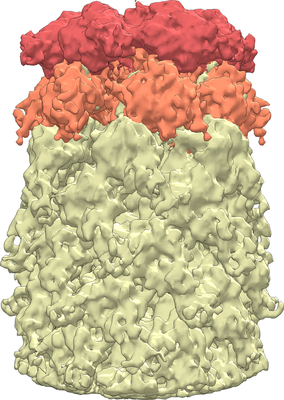 emd_3878.png emd_3878.png | 173 KB | ||
| Masks | emd_3878_msk_1.map | 64 MB | Mask map | |
| Others | emd_3878_additional.map.gzemd_3878_additional_1.map.gzemd_3878_half_map_1.map.gzemd_3878_half_map_2.map.gz | 59.6 MB 59.6 MB 48.4 MB 48.4 MB | ||
| Archive directory |  http://ftp.pdbj.org/pub/emdb/structures/EMD-3878ftp://ftp.pdbj.org/pub/emdb/structures/EMD-3878 http://ftp.pdbj.org/pub/emdb/structures/EMD-3878ftp://ftp.pdbj.org/pub/emdb/structures/EMD-3878 | HTTPS FTP |
-Related structure data


-Links
| EMDB pages | EMDB (EBI/PDBe) / EMDataResource |
|---|
-Map
| File | Download / File: emd_3878.map.gz / Format: CCP4 / Size: 64 MB / Type: IMAGE STORED AS FLOATING POINT NUMBER (4 BYTES) | ||||||||||||||||||||||||||||||||||||||||||||||||||||||||||||||||||||
|---|---|---|---|---|---|---|---|---|---|---|---|---|---|---|---|---|---|---|---|---|---|---|---|---|---|---|---|---|---|---|---|---|---|---|---|---|---|---|---|---|---|---|---|---|---|---|---|---|---|---|---|---|---|---|---|---|---|---|---|---|---|---|---|---|---|---|---|---|---|
| Annotation | The distal end of a non-contractile T6SS sheath. | ||||||||||||||||||||||||||||||||||||||||||||||||||||||||||||||||||||
| Voxel size | X=Y=Z: 2.12 Å | ||||||||||||||||||||||||||||||||||||||||||||||||||||||||||||||||||||
| Density |
| ||||||||||||||||||||||||||||||||||||||||||||||||||||||||||||||||||||
| Symmetry | Space group: 1 | ||||||||||||||||||||||||||||||||||||||||||||||||||||||||||||||||||||
| Details | EMDB XML:
CCP4 map header:
| ||||||||||||||||||||||||||||||||||||||||||||||||||||||||||||||||||||
-Supplemental data
-Mask #1
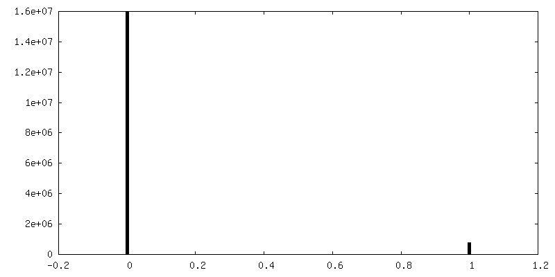
| File | emd_3878_msk_1.map | ||||||||||||
|---|---|---|---|---|---|---|---|---|---|---|---|---|---|
| Projections & Slices |
| ||||||||||||
| Density Histograms |
 Z
Z Y
Y X
X


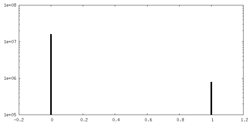
-Additional map: The distal end of a non-contractile T6SS sheath. Post-processed map.
| File | emd_3878_additional.map | ||||||||||||
|---|---|---|---|---|---|---|---|---|---|---|---|---|---|
| Annotation | The distal end of a non-contractile T6SS sheath. Post-processed map. | ||||||||||||
| Projections & Slices |
| ||||||||||||
| Density Histograms |


-Additional map: The distal end of a non-contractile T6SS sheath. Post-processed map.
| File | emd_3878_additional_1.map | ||||||||||||
|---|---|---|---|---|---|---|---|---|---|---|---|---|---|
| Annotation | The distal end of a non-contractile T6SS sheath. Post-processed map. | ||||||||||||
| Projections & Slices |
| ||||||||||||
| Density Histograms |


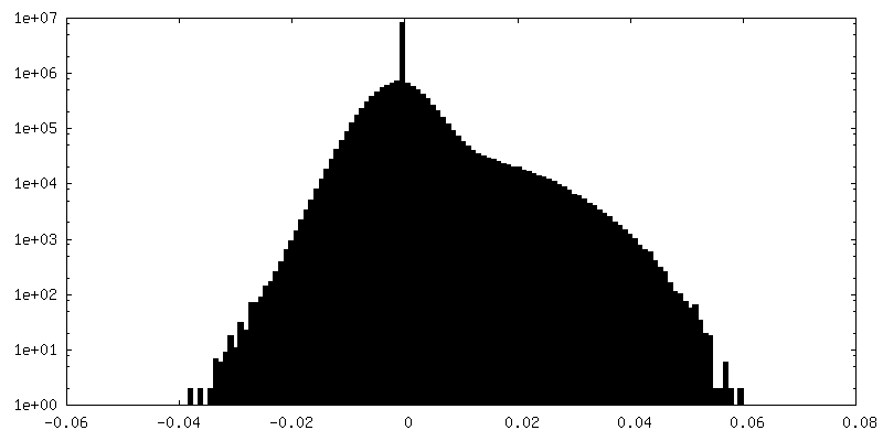
-Half map: #1
| File | emd_3878_half_map_1.map | ||||||||||||
|---|---|---|---|---|---|---|---|---|---|---|---|---|---|
| Projections & Slices |
| ||||||||||||
| Density Histograms |


-Half map: #2
| File | emd_3878_half_map_2.map | ||||||||||||
|---|---|---|---|---|---|---|---|---|---|---|---|---|---|
| Projections & Slices |
| ||||||||||||
| Density Histograms |


- Sample components
-Entire : The distal end of a non-contractile T6SS sheath.
| Entire | Name: The distal end of a non-contractile T6SS sheath. |
|---|---|
| Components |
|
-Supramolecule #1: The distal end of a non-contractile T6SS sheath.
| Supramolecule | Name: The distal end of a non-contractile T6SS sheath. / type: organelle_or_cellular_component / ID: 1 / Parent: 0 |
|---|---|
| Source (natural) | Organism: Vibrio cholerae (bacteria) / Strain: 2740-80 / Organelle: T6SS |
| Molecular weight | Experimental: 540 KDa |
-Experimental details
-Structure determination
| Method | cryo EM |
|---|---|
 Processing Processing | single particle reconstruction |
| Aggregation state | filament |
-Sample preparation
| Buffer | pH: 8.3 |
|---|---|
| Grid | Model: Quantifoil R2/1 / Material: COPPER / Mesh: 400 / Support film - Material: CARBON / Support film - topology: HOLEY / Pretreatment - Type: GLOW DISCHARGE / Pretreatment - Atmosphere: AIR |
| Vitrification | Cryogen name: ETHANE / Chamber temperature: 277 K / Instrument: FEI VITROBOT MARK IV / Details: blot for 3 seconds before plunging. |
- Electron microscopy
| Microscope | FEI TITAN KRIOS |
|---|---|
| Electron beam | Acceleration voltage: 300 kV / Electron source: FIELD EMISSION GUN |
| Electron optics | Illumination mode: FLOOD BEAM / Imaging mode: BRIGHT FIELDBright-field microscopy / Cs: 2.7 mm / Nominal defocus max: 3.0 µm / Nominal defocus min: 1.5 µm / Nominal magnification: 130000 |
| Specialist optics | Energy filter - Name: GIF Quantum LS |
| Sample stage | Specimen holder model: FEI TITAN KRIOS AUTOGRID HOLDER / Cooling holder cryogen: NITROGEN |
| Image recording | Film or detector model: GATAN K2 SUMMIT (4k x 4k) / Detector mode: COUNTING / Number real images: 9202 / Average exposure time: 16.0 sec. / Average electron dose: 5.0 e/Å2 |
| Experimental equipment |  Model: Titan Krios / Image courtesy: FEI Company |
-Image processing
| Particle selection | Number selected: 21446 |
|---|---|
| CTF correction | Software - Name: Gctf (ver. 1.06) |
| Initial angle assignment | Type: OTHER / Software - Name: RELION (ver. 1.4) / Details: Probability-weighted angular assignment |
| Final angle assignment | Type: OTHER / Software - Name: RELION (ver. 2.1) / Details: Probability-weighted angular assignment |
| Final reconstruction | Applied symmetry - Point group: C6 (6 fold cyclic) / Resolution.type: BY AUTHOR / Resolution: 7.5 Å / Resolution method: FSC 0.143 CUT-OFF / Software - Name: RELION (ver. 2.1) / Number images used: 3710 |
| FSC plot (resolution estimation) | 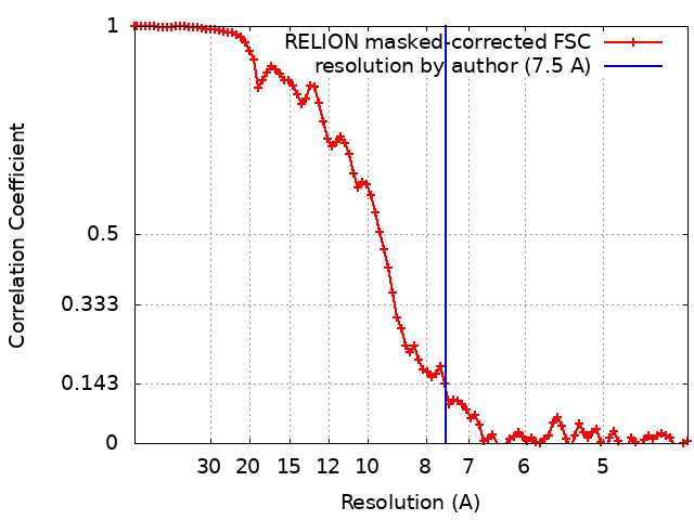 |
