 Movie
Movie Controller


+ Open data

- Basic information
| Entry | Database: EMDB / ID: EMD-20617 | |||||||||
|---|---|---|---|---|---|---|---|---|---|---|
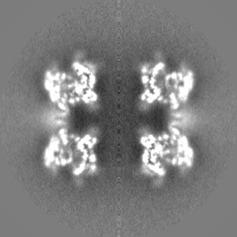
| Title | EM map of MPEG-1 (w.t.) soluble pre-pore complex | |||||||||
 Map data Map data | MPEG-1 (w.t.) soluble pre-pore complex, sharpened map | |||||||||
 Sample Sample |
| |||||||||
| Biological species |   Homo sapiens (human) Homo sapiens (human) | |||||||||
| Method | single particle reconstruction / cryo EM / Resolution: 3.5 Å | |||||||||
 Authors Authors | Pang SS / Bayly-Jones C | |||||||||
| Funding support |  Australia, 1 items Australia, 1 items
| |||||||||
 Citation Citation | Journal: Nat Commun / Year: 2019 Title: The cryo-EM structure of the acid activatable pore-forming immune effector Macrophage-expressed gene 1. Authors: Siew Siew Pang / Charles Bayly-Jones / Mazdak Radjainia / Bradley A Spicer / Ruby H P Law / Adrian W Hodel / Edward S Parsons / Susan M Ekkel / Paul J Conroy / Georg Ramm / Hariprasad ...Authors: Siew Siew Pang / Charles Bayly-Jones / Mazdak Radjainia / Bradley A Spicer / Ruby H P Law / Adrian W Hodel / Edward S Parsons / Susan M Ekkel / Paul J Conroy / Georg Ramm / Hariprasad Venugopal / Phillip I Bird / Bart W Hoogenboom / Ilia Voskoboinik / Yann Gambin / Emma Sierecki / Michelle A Dunstone / James C Whisstock /   Abstract: Macrophage-expressed gene 1 (MPEG1/Perforin-2) is a perforin-like protein that functions within the phagolysosome to damage engulfed microbes. MPEG1 is thought to form pores in target membranes, ...Macrophage-expressed gene 1 (MPEG1/Perforin-2) is a perforin-like protein that functions within the phagolysosome to damage engulfed microbes. MPEG1 is thought to form pores in target membranes, however, its mode of action remains unknown. We use cryo-Electron Microscopy (cryo-EM) to determine the 2.4 Å structure of a hexadecameric assembly of MPEG1 that displays the expected features of a soluble prepore complex. We further discover that MPEG1 prepore-like assemblies can be induced to perforate membranes through acidification, such as would occur within maturing phagolysosomes. We next solve the 3.6 Å cryo-EM structure of MPEG1 in complex with liposomes. These data reveal that a multi-vesicular body of 12 kDa (MVB12)-associated β-prism (MABP) domain binds membranes such that the pore-forming machinery of MPEG1 is oriented away from the bound membrane. This unexpected mechanism of membrane interaction suggests that MPEG1 remains bound to the phagolysosome membrane while simultaneously forming pores in engulfed bacterial targets. | |||||||||
| History |
|
- Structure visualization
| Movie |
 Movie viewer Movie viewer |
|---|---|
| Structure viewer | EM map: SurfViewMolmilJmol/JSmol |
| Supplemental images |

- Downloads & links
-EMDB archive
| Map data | emd_20617.map.gz | 28.6 MB | EMDB map data format | |
|---|---|---|---|---|
| Header (meta data) | emd-20617-v30.xmlemd-20617.xml | 16.7 KB 16.7 KB | Display Display | EMDB header |
| FSC (resolution estimation) | emd_20617_fsc.xml | 11.9 KB | Display | FSC data file |
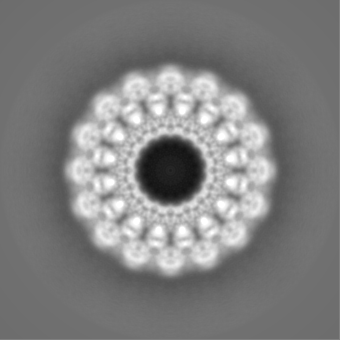
| Images | 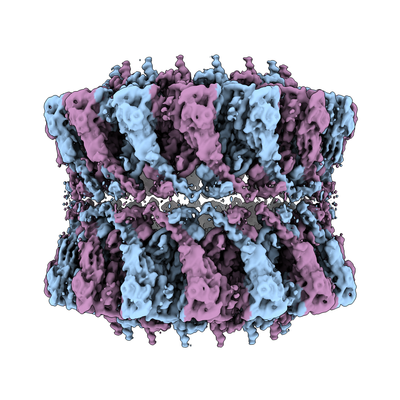 emd_20617.png emd_20617.png | 179.5 KB | ||
| Masks | emd_20617_msk_1.map | 149.9 MB | Mask map | |
| Others | emd_20617_additional.map.gzemd_20617_additional_1.map.gzemd_20617_half_map_1.map.gzemd_20617_half_map_2.map.gz | 110.9 MB 110.9 MB 111.4 MB 111.4 MB | ||
| Archive directory |  http://ftp.pdbj.org/pub/emdb/structures/EMD-20617ftp://ftp.pdbj.org/pub/emdb/structures/EMD-20617 http://ftp.pdbj.org/pub/emdb/structures/EMD-20617ftp://ftp.pdbj.org/pub/emdb/structures/EMD-20617 | HTTPS FTP |
-Related structure data


-Links
| EMDB pages | EMDB (EBI/PDBe) / EMDataResource |
|---|
-Map
| File | Download / File: emd_20617.map.gz / Format: CCP4 / Size: 149.9 MB / Type: IMAGE STORED AS FLOATING POINT NUMBER (4 BYTES) | ||||||||||||||||||||||||||||||||||||||||||||||||||||||||||||||||||||
|---|---|---|---|---|---|---|---|---|---|---|---|---|---|---|---|---|---|---|---|---|---|---|---|---|---|---|---|---|---|---|---|---|---|---|---|---|---|---|---|---|---|---|---|---|---|---|---|---|---|---|---|---|---|---|---|---|---|---|---|---|---|---|---|---|---|---|---|---|---|
| Annotation | MPEG-1 (w.t.) soluble pre-pore complex, sharpened map | ||||||||||||||||||||||||||||||||||||||||||||||||||||||||||||||||||||
| Voxel size | X=Y=Z: 1.1 Å | ||||||||||||||||||||||||||||||||||||||||||||||||||||||||||||||||||||
| Density |
| ||||||||||||||||||||||||||||||||||||||||||||||||||||||||||||||||||||
| Symmetry | Space group: 1 | ||||||||||||||||||||||||||||||||||||||||||||||||||||||||||||||||||||
| Details | EMDB XML:
CCP4 map header:
| ||||||||||||||||||||||||||||||||||||||||||||||||||||||||||||||||||||
-Supplemental data
-Mask #1
| File | emd_20617_msk_1.map | ||||||||||||
|---|---|---|---|---|---|---|---|---|---|---|---|---|---|
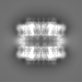
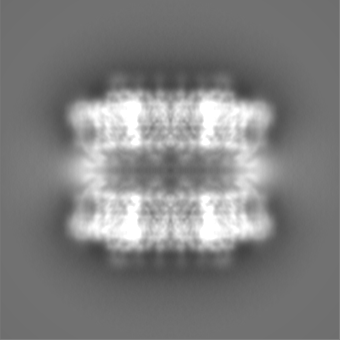
| Projections & Slices |
| ||||||||||||
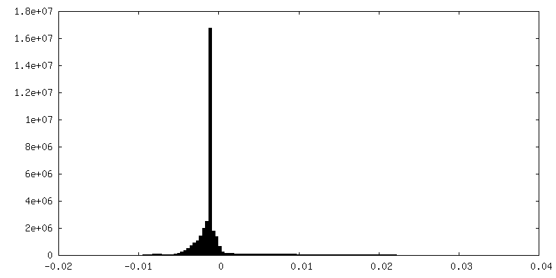
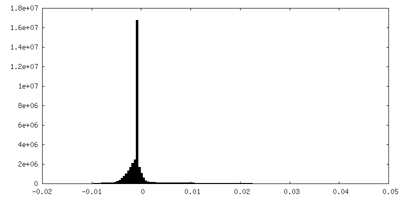
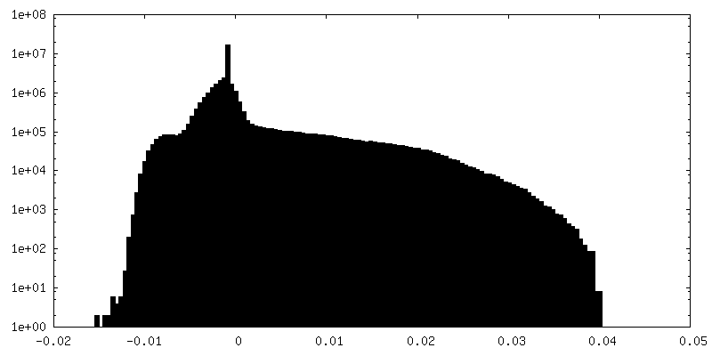
| Density Histograms |
 Z
Z Y
Y X
X


-Additional map: MPEG-1 (w.t.) soluble pre-pore complex, unsharpened map
| File | emd_20617_additional.map | ||||||||||||
|---|---|---|---|---|---|---|---|---|---|---|---|---|---|
| Annotation | MPEG-1 (w.t.) soluble pre-pore complex, unsharpened map | ||||||||||||
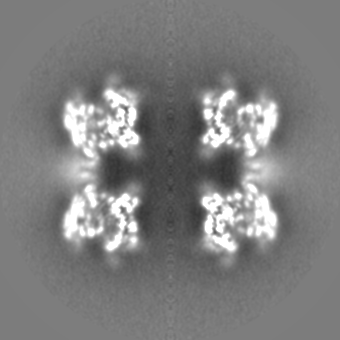
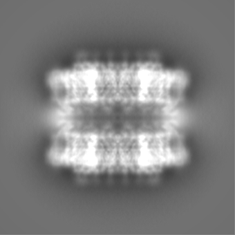
| Projections & Slices |
| ||||||||||||
| Density Histograms |


-Additional map: MPEG-1 (w.t.) soluble pre-pore complex, unsharpened map
| File | emd_20617_additional_1.map | ||||||||||||
|---|---|---|---|---|---|---|---|---|---|---|---|---|---|
| Annotation | MPEG-1 (w.t.) soluble pre-pore complex, unsharpened map | ||||||||||||
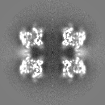
| Projections & Slices |
| ||||||||||||
| Density Histograms |


-Half map: MPEG-1 (w.t.) soluble pre-pore complex, half map #1
| File | emd_20617_half_map_1.map | ||||||||||||
|---|---|---|---|---|---|---|---|---|---|---|---|---|---|
| Annotation | MPEG-1 (w.t.) soluble pre-pore complex, half map #1 | ||||||||||||
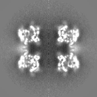
| Projections & Slices |
| ||||||||||||
| Density Histograms |


-Half map: MPEG-1 (w.t.) soluble pre-pore complex, half map #2
| File | emd_20617_half_map_2.map | ||||||||||||
|---|---|---|---|---|---|---|---|---|---|---|---|---|---|
| Annotation | MPEG-1 (w.t.) soluble pre-pore complex, half map #2 | ||||||||||||
| Projections & Slices |
| ||||||||||||
| Density Histograms |

- Sample components
-Entire : Head-to-head assembly of MPEG-1 (w.t.) soluble pre-pore
| Entire | Name: Head-to-head assembly of MPEG-1 (w.t.) soluble pre-pore |
|---|---|
| Components |
|
-Supramolecule #1: Head-to-head assembly of MPEG-1 (w.t.) soluble pre-pore
| Supramolecule | Name: Head-to-head assembly of MPEG-1 (w.t.) soluble pre-pore type: complex / ID: 1 / Parent: 0 / Macromolecule list: all |
|---|---|
| Source (natural) | Organism: Homo sapiens (human) |
| Recombinant expression | Organism:   Spodoptera frugiperda (fall armyworm) Spodoptera frugiperda (fall armyworm) |
| Molecular weight | Theoretical: 2.3 MDa |
-Macromolecule #1: Macrophage-Expressed Gene 1 protein (MPEG-1)
| Macromolecule | Name: Macrophage-Expressed Gene 1 protein (MPEG-1) / type: protein_or_peptide / ID: 1 / Enantiomer: LEVO |
|---|---|
| Source (natural) | Organism: Homo sapiens (human) |
| Recombinant expression | Organism: Spodoptera frugiperda (fall armyworm) |
| Sequence | String: KSGKPSGEMD EVGVQKCKNA LKLPVLEVLP GGGWDNLRNV DMGRVMELTY SNCRTTEDGQ YIIPDEIFTI PQKQSNLEMN SEILESWANY QSSTSYSINT ELSLFSKVNG KFSTEFQRMK TLQVKDQAIT TRVQVRNLVY TVKINPTLEL SSGFRKELLD ISDRLENNQT ...String: KSGKPSGEMD EVGVQKCKNA LKLPVLEVLP GGGWDNLRNV DMGRVMELTY SNCRTTEDGQ YIIPDEIFTI PQKQSNLEMN SEILESWANY QSSTSYSINT ELSLFSKVNG KFSTEFQRMK TLQVKDQAIT TRVQVRNLVY TVKINPTLEL SSGFRKELLD ISDRLENNQT RMATYLAELL VLNYGTHVTT SVDAGAALIQ EDHLRASFLQ DSQSSRSAVT ASAGLAFQNT VNFKFEENYT SQNVLTKSYL SNRTNSRVQS IGGVPFYPGI TLQAWQQGIT NHLVAIDRSG LPLHFFINPN MLPDLPGPLV KKVSKTVETA VKRYYTFNTY PGCTDLNSPN FNFQANTDDG SCEGKMTNFS FGGVYQECTQ LSGNRDVLLC QKLEQKNPLT GDFSCPSGYS PVHLLSQIHE EGYNHLECHR KCTLLVFCKT VCEDVFQVAK AEFRAFWCVA SSQVPENSGL LFGGLFSSKS INPMTNAQSC PAGYFPLRLF ENLKVCVSQD YELGSRFAVP FGGFFSCTVG NPLVDPAISR DLGAPSLKKC PGGFSQHPAL ISDGCQVSYC VKSGLFTGGS LPPARLPPFT RPPLMSQAAT NTVIVTNSEN ARSWIKDSQT HQWRLGEPIE LRRAMNVIHG DGGGLSHHHH HH |
-Experimental details
-Structure determination
| Method | cryo EM |
|---|---|
 Processing Processing | single particle reconstruction |
| Aggregation state | particle |
-Sample preparation
| Concentration | 2 mg/mL |
|---|---|
| Buffer | pH: 7.2 |
| Grid | Details: unspecified |
| Vitrification | Cryogen name: ETHANE |
- Electron microscopy
| Microscope | FEI TITAN KRIOS |
|---|---|
| Electron beam | Acceleration voltage: 300 kV / Electron source: FIELD EMISSION GUN |
| Electron optics | Illumination mode: FLOOD BEAM / Imaging mode: BRIGHT FIELDBright-field microscopy |
| Image recording | Film or detector model: FEI FALCON II (4k x 4k) / Average electron dose: 45.0 e/Å2 |
| Experimental equipment |  Model: Titan Krios / Image courtesy: FEI Company |
-Image processing
| Initial angle assignment | Type: NOT APPLICABLE |
|---|---|
| Final angle assignment | Type: NOT APPLICABLE |
| Final reconstruction | Applied symmetry - Point group: D16 (2x16 fold dihedral) / Resolution.type: BY AUTHOR / Resolution: 3.5 Å / Resolution method: FSC 0.143 CUT-OFF / Software - Name: RELION (ver. 2.1) / Number images used: 25359 |
| FSC plot (resolution estimation) | 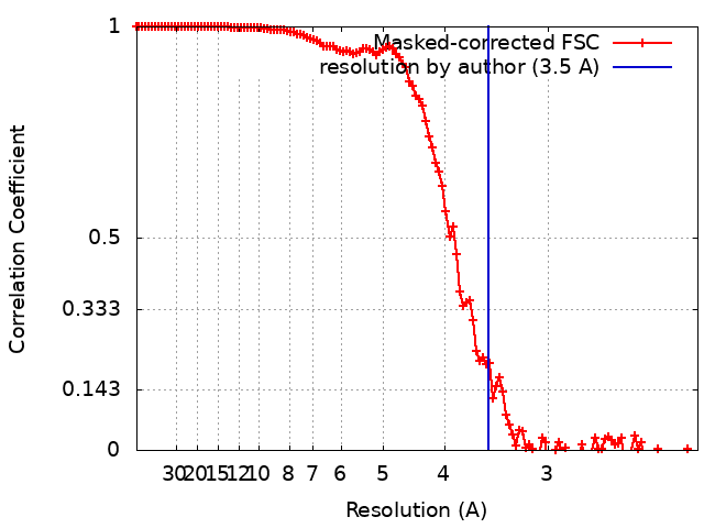 |
