ジャーナル: Mol Cell / 年: 2024 タイトル: Structure of the multi-subunit chloroplast RNA polymerase. 著者: Paula F V do Prado / Frederik M Ahrens / Monique Liebers / Noah Ditz / Hans-Peter Braun / Thomas Pfannschmidt / Hauke S Hillen / 要旨: Chloroplasts contain a dedicated genome that encodes subunits of the photosynthesis machinery. Transcription of photosynthesis genes is predominantly carried out by a plastid-encoded RNA polymerase ...Chloroplasts contain a dedicated genome that encodes subunits of the photosynthesis machinery. Transcription of photosynthesis genes is predominantly carried out by a plastid-encoded RNA polymerase (PEP), a nearly 1 MDa complex composed of core subunits with homology to eubacterial RNA polymerases (RNAPs) and at least 12 additional chloroplast-specific PEP-associated proteins (PAPs). However, the architecture of this complex and the functions of the PAPs remain unknown. Here, we report the cryo-EM structure of a 19-subunit PEP complex from Sinapis alba (white mustard). The structure reveals that the PEP core resembles prokaryotic and nuclear RNAPs but contains chloroplast-specific features that mediate interactions with the PAPs. The PAPs are unrelated to known transcription factors and arrange around the core in a unique fashion. Their structures suggest potential functions during transcription in the chemical environment of chloroplasts. These results reveal structural insights into chloroplast transcription and provide a framework for understanding photosynthesis gene expression.
 ムービー
ムービー コントローラー


 データを開く
 基本情報
基本情報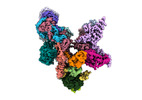
 マップデータ
 試料
試料 キーワード
キーワード 機能・相同性情報
 Sinapis alba (シロガラシ)
 データ登録者
 ドイツ, 5件
  引用
引用 構造の表示
構造の表示
 ダウンロードとリンク
 emd_18496.png
 http://ftp.pdbj.org/pub/emdb/structures/EMD-18496


 試料の構成要素
 解析
解析 電子顕微鏡法
 FIELD EMISSION GUN
