 Movie
Movie Controller

[English] 日本語
 Yorodumi
- EMDB-1059: Electron cryomicroscopy structure of N-ethyl maleimide sensitive ... -
+ Open data

- Basic information
| Entry | Database: EMDB / ID: EMD-1059 | |||||||||
|---|---|---|---|---|---|---|---|---|---|---|
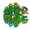
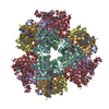
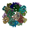
| Title | Electron cryomicroscopy structure of N-ethyl maleimide sensitive factor at 11 A resolution. | |||||||||
 Map data Map data | 3D density map of NSF?alpha-SNAP?SNARE (20S) | |||||||||
 Sample Sample |
| |||||||||
| Biological species |    Cricetulus griseus (Chinese hamster) / Bos taurus (cattle) / unidentified (others) Cricetulus griseus (Chinese hamster) / Bos taurus (cattle) / unidentified (others) | |||||||||
| Method | single particle reconstruction / cryo EM / negative staining / Resolution: 11.0 Å | |||||||||
 Authors Authors | Furst J / Sutton RB / Chen J / Brunger AT / Grigorieff N | |||||||||
 Citation Citation | Journal: EMBO J / Year: 2003 Title: Electron cryomicroscopy structure of N-ethyl maleimide sensitive factor at 11 A resolution. Authors: Johannes Furst / R Bryan Sutton / James Chen / Axel T Brunger / Nikolaus Grigorieff /  Abstract: N-ethyl maleimide sensitive factor (NSF) belongs to the AAA family of ATPases and is involved in a number of cellular functions, including vesicle fusion and trafficking of membrane proteins. We ...N-ethyl maleimide sensitive factor (NSF) belongs to the AAA family of ATPases and is involved in a number of cellular functions, including vesicle fusion and trafficking of membrane proteins. We present the three-dimensional structure of the hydrolysis mutant E329Q of NSF complexed with an ATP-ADP mixture at 11 A resolution by electron cryomicroscopy and single-particle averaging of NSF.alpha-SNAP.SNARE complexes. The NSF domains D1 and D2 form hexameric rings that are arranged in a double-layered barrel. Our structure is more consistent with an antiparallel orientation of the two rings rather than a parallel one. The crystal structure of the D2 domain of NSF was docked into the EM density map and shows good agreement, including details at the secondary structural level. Six protrusions corresponding to the N domain of NSF (NSF-N) emerge from the sides of the D1 domain ring. The density corresponding to alpha-SNAP and SNAREs is located on the 6-fold axis of the structure, near the NSF-N domains. The density of the N domain is weak, suggesting conformational variability in this part of NSF. | |||||||||
| History |
|
- Structure visualization
| Movie |
 Movie viewer Movie viewer |
|---|---|
| Structure viewer | EM map: SurfViewMolmilJmol/JSmol |
| Supplemental images |

- Downloads & links
-EMDB archive
| Map data | emd_1059.map.gz | 1.7 MB | EMDB map data format | |
|---|---|---|---|---|
| Header (meta data) | emd-1059-v30.xmlemd-1059.xml | 10.5 KB 10.5 KB | Display Display | EMDB header |
| Images | 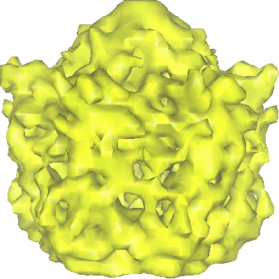 1059.gif 1059.gif | 76.8 KB | ||
| Archive directory |  http://ftp.pdbj.org/pub/emdb/structures/EMD-1059ftp://ftp.pdbj.org/pub/emdb/structures/EMD-1059 http://ftp.pdbj.org/pub/emdb/structures/EMD-1059ftp://ftp.pdbj.org/pub/emdb/structures/EMD-1059 | HTTPS FTP |
-Related structure data
| Similar structure data |
|---|
-Links
| EMDB pages | EMDB (EBI/PDBe) / EMDataResource |
|---|
-Map
| File | Download / File: emd_1059.map.gz / Format: CCP4 / Size: 2.7 MB / Type: IMAGE STORED AS FLOATING POINT NUMBER (4 BYTES) | ||||||||||||||||||||||||||||||||||||||||||||||||||||||||||||||||||||
|---|---|---|---|---|---|---|---|---|---|---|---|---|---|---|---|---|---|---|---|---|---|---|---|---|---|---|---|---|---|---|---|---|---|---|---|---|---|---|---|---|---|---|---|---|---|---|---|---|---|---|---|---|---|---|---|---|---|---|---|---|---|---|---|---|---|---|---|---|---|
| Annotation | 3D density map of NSF?alpha-SNAP?SNARE (20S) | ||||||||||||||||||||||||||||||||||||||||||||||||||||||||||||||||||||
| Voxel size | X=Y=Z: 3.5 Å | ||||||||||||||||||||||||||||||||||||||||||||||||||||||||||||||||||||
| Density |
| ||||||||||||||||||||||||||||||||||||||||||||||||||||||||||||||||||||
| Symmetry | Space group: 1 | ||||||||||||||||||||||||||||||||||||||||||||||||||||||||||||||||||||
| Details | EMDB XML:
CCP4 map header:
| ||||||||||||||||||||||||||||||||||||||||||||||||||||||||||||||||||||
-Supplemental data
- Sample components
-Entire : NSF alpha-SNAP SNARE 20S
| Entire | Name: NSF alpha-SNAP SNARE 20S |
|---|---|
| Components |
|
-Supramolecule #1000: NSF alpha-SNAP SNARE 20S
| Supramolecule | Name: NSF alpha-SNAP SNARE 20S / type: sample / ID: 1000 Details: The sample was monodisperse. Some domains were disordered. Number unique components: 3 |
|---|---|
| Molecular weight | Experimental: 680 KDa / Method: gel permeation chromatography |
-Macromolecule #1: N-ethyl Maleimide Sensitive Factor
| Macromolecule | Name: N-ethyl Maleimide Sensitive Factor / type: protein_or_peptide / ID: 1 / Name.synonym: NSF / Details: 6 subunits / Number of copies: 6 / Oligomeric state: hexamer / Recombinant expression: Yes |
|---|---|
| Source (natural) | Organism: Cricetulus griseus (Chinese hamster) / synonym: Chinese Hamster |
| Molecular weight | Experimental: 82.8 KDa |
| Recombinant expression | Organism:  Escherichia coli (E. coli) Escherichia coli (E. coli) |
-Macromolecule #2: Soluble NSF Attachment Protein
| Macromolecule | Name: Soluble NSF Attachment Protein / type: protein_or_peptide / ID: 2 / Name.synonym: alpha-SNAP / Recombinant expression: Yes |
|---|---|
| Source (natural) | Organism: Bos taurus (cattle) / synonym: Cattle |
| Molecular weight | Experimental: 33.4 KDa |
-Macromolecule #3: Soluble NSF Attachment Protein Receptor
| Macromolecule | Name: Soluble NSF Attachment Protein Receptor / type: protein_or_peptide / ID: 3 / Name.synonym: SNARE / Recombinant expression: Yes |
|---|---|
| Source (natural) | Organism: unidentified (others) / synonym: Chinese Hamster |
| Molecular weight | Experimental: 82.3 KDa |
-Experimental details
-Structure determination
| Method | negative staining, cryo EM |
|---|---|
 Processing Processing | single particle reconstruction |
| Aggregation state | particle |
-Sample preparation
| Concentration | 0.2 mg/mL |
|---|---|
| Buffer | pH: 7.4 Details: 20mM HEPES, pH 7.4, 300 mM NACl, 2 mM MgCl2, 5mM b-mercaptoethanol, 0.01/0.1 mM ADP/ATP nucleotide |
| Staining | Type: NEGATIVE / Details: cryo (no stain) |
| Grid | Details: holey carbon film |
| Vitrification | Cryogen name: ETHANE / Chamber humidity: 80 % / Chamber temperature: 85 K / Instrument: HOMEMADE PLUNGER / Details: Vitrification instrument: Technion plunger |
- Electron microscopy
| Microscope | FEI TECNAI 20 |
|---|---|
| Electron beam | Acceleration voltage: 200 kV / Electron source: FIELD EMISSION GUN |
| Electron optics | Illumination mode: FLOOD BEAM / Imaging mode: BRIGHT FIELDBright-field microscopy / Cs: 2.0 mm / Nominal defocus max: 4.0 µm / Nominal defocus min: 2.0 µm / Nominal magnification: 62000 |
| Sample stage | Specimen holder: Eucentric / Specimen holder model: GATAN LIQUID NITROGEN |
| Temperature | Average: 85 K |
| Image recording | Category: FILM / Film or detector model: KODAK SO-163 FILM / Digitization - Scanner: ZEISS SCAI / Digitization - Sampling interval: 7 µm / Average electron dose: 10 e/Å2 / Details: 3 x 3 averaging, final step size = 27 microns / Od range: 1.4 / Bits/pixel: 8 |
| Tilt angle min | 0 |
| Tilt angle max | 0 |
-Image processing
| CTF correction | Details: FREALIGN |
|---|---|
| Final reconstruction | Applied symmetry - Point group: C6 (6 fold cyclic) / Algorithm: OTHER / Resolution.type: BY AUTHOR / Resolution: 11.0 Å / Resolution method: FSC 0.5 CUT-OFF / Software - Name: IMAGIC, FREALIGN / Details: Final map was calculated from three data sets / Number images used: 31592 |
| Details | manual particle selection |
