 Movie
Movie Controller


+ Open data

- Basic information
| Entry | Database: EMDB / ID: EMD-0622 | |||||||||
|---|---|---|---|---|---|---|---|---|---|---|
| Title | Structure of the AAV2 with its Cell Receptor, AAVR | |||||||||
 Map data Map data | AAV2 bound with AAVR (class2) | |||||||||
 Sample Sample |
| |||||||||
| Biological species |    Adeno-associated virus Adeno-associated virus | |||||||||
| Method | subtomogram averaging / cryo EM / Resolution: 20.0 Å | |||||||||
 Authors Authors | Hu GQ / Meyer NL / Stagg SM / Chapman MS / Davulcu O / Xie Q / Noble AJ / Yoshioka C / Gingerich D / Trzynka A / David L | |||||||||
 Citation Citation | Journal: Elife / Year: 2019 Title: Structure of the gene therapy vector, adeno-associated virus with its cell receptor, AAVR. Authors: Nancy L Meyer / Guiqing Hu / Omar Davulcu / Qing Xie / Alex J Noble / Craig Yoshioka / Drew S Gingerich / Andrew Trzynka / Larry David / Scott M Stagg / Michael Stewart Chapman /  Abstract: Adeno-associated virus (AAV) vectors are preeminent in emerging clinical gene therapies. Generalizing beyond the most tractable genetic diseases will require modulation of cell specificity and immune ...Adeno-associated virus (AAV) vectors are preeminent in emerging clinical gene therapies. Generalizing beyond the most tractable genetic diseases will require modulation of cell specificity and immune neutralization. Interactions of AAV with its cellular receptor, AAVR, are key to understanding cell-entry and trafficking with the rigor needed to engineer tissue-specific vectors. -electron tomography shows ordered binding of part of the flexible receptor to the viral surface, with distal domains in multiple conformations. Regions of the virus and receptor in close physical proximity can be identified by cross-linking/mass spectrometry. -electron microscopy with a two-domain receptor fragment reveals the interactions at 2.4 Å resolution. AAVR binds between AAV's spikes on a plateau that is conserved, except in one clade whose structure is AAVR-incompatible. AAVR's footprint overlaps the epitopes of several neutralizing antibodies, prompting a re-evaluation of neutralization mechanisms. The structure provides a roadmap for experimental probing and manipulation of viral-receptor interactions. | |||||||||
| History |
|
- Structure visualization
| Movie |
 Movie viewer Movie viewer |
|---|---|
| Structure viewer | EM map: SurfViewMolmilJmol/JSmol |
| Supplemental images |

- Downloads & links
-EMDB archive
| Map data | emd_0622.map.gz | 136.4 KB | EMDB map data format | |
|---|---|---|---|---|
| Header (meta data) | emd-0622-v30.xmlemd-0622.xml | 14.5 KB 14.5 KB | Display Display | EMDB header |
| Images | 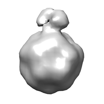 emd_0622.png emd_0622.png | 46 KB | ||
| Archive directory |  http://ftp.pdbj.org/pub/emdb/structures/EMD-0622ftp://ftp.pdbj.org/pub/emdb/structures/EMD-0622 http://ftp.pdbj.org/pub/emdb/structures/EMD-0622ftp://ftp.pdbj.org/pub/emdb/structures/EMD-0622 | HTTPS FTP |
-Related structure data


-Links
| EMDB pages | EMDB (EBI/PDBe) / EMDataResource |
|---|
-Map
| File | Download / File: emd_0622.map.gz / Format: CCP4 / Size: 182.6 KB / Type: IMAGE STORED AS FLOATING POINT NUMBER (4 BYTES) | ||||||||||||||||||||||||||||||||||||||||||||||||||||||||||||
|---|---|---|---|---|---|---|---|---|---|---|---|---|---|---|---|---|---|---|---|---|---|---|---|---|---|---|---|---|---|---|---|---|---|---|---|---|---|---|---|---|---|---|---|---|---|---|---|---|---|---|---|---|---|---|---|---|---|---|---|---|---|
| Annotation | AAV2 bound with AAVR (class2) | ||||||||||||||||||||||||||||||||||||||||||||||||||||||||||||
| Voxel size | X=Y=Z: 15.6 Å | ||||||||||||||||||||||||||||||||||||||||||||||||||||||||||||
| Density |
| ||||||||||||||||||||||||||||||||||||||||||||||||||||||||||||
| Symmetry | Space group: 1 | ||||||||||||||||||||||||||||||||||||||||||||||||||||||||||||
| Details | EMDB XML:
CCP4 map header:
| ||||||||||||||||||||||||||||||||||||||||||||||||||||||||||||
-Supplemental data
- Sample components
-Entire : AAV2 complex with AAVR receptor
| Entire | Name: AAV2 complex with AAVR receptor |
|---|---|
| Components |
|
-Supramolecule #1: AAV2 complex with AAVR receptor
| Supramolecule | Name: AAV2 complex with AAVR receptor / type: complex / ID: 1 / Parent: 0 / Details: class 1 |
|---|---|
| Source (natural) | Organism: Adeno-associated virus / Strain: hybrid of serotypes 2, 8, and 9 |
| Recombinant expression | Organism:   Spodoptera frugiperda (fall armyworm) / Recombinant strain: SF9 / Recombinant cell: Sf9 / Recombinant plasmid: pFBDDJM11 Spodoptera frugiperda (fall armyworm) / Recombinant strain: SF9 / Recombinant cell: Sf9 / Recombinant plasmid: pFBDDJM11 |
| Molecular weight | Theoretical: 3.75 MDa |
-Experimental details
-Structure determination
| Method | cryo EM |
|---|---|
 Processing Processing | subtomogram averaging |
| Aggregation state | particle |
-Sample preparation
| Concentration | 0.6 mg/mL | ||||||||||||
|---|---|---|---|---|---|---|---|---|---|---|---|---|---|
| Buffer | pH: 7.4 Component:
Details: blot force = 1, blot time = 3 seconds, total blots = 1 | ||||||||||||
| Vitrification | Cryogen name: ETHANE / Chamber humidity: 100 % / Chamber temperature: 4 K / Instrument: FEI VITROBOT MARK IV |
- Electron microscopy
| Microscope | FEI TITAN KRIOS |
|---|---|
| Electron beam | Acceleration voltage: 300 kV / Electron source: FIELD EMISSION GUN |
| Electron optics | C2 aperture diameter: 70.0 µm / Calibrated defocus max: 0.011 µm / Calibrated defocus min: 9.0 µm / Calibrated magnification: 18000 / Illumination mode: FLOOD BEAM / Imaging mode: BRIGHT FIELDBright-field microscopy / Cs: 2.7 mm / Nominal defocus max: 0.011 µm / Nominal defocus min: 9.0 µm / Nominal magnification: 18000 |
| Sample stage | Specimen holder model: FEI TITAN KRIOS AUTOGRID HOLDER / Cooling holder cryogen: NITROGEN |
| Temperature | Min: 290.0 K / Max: 300.0 K |
| Details | preliminary grid screening was performed manually |
| Image recording | Film or detector model: DIRECT ELECTRON DE-20 (5k x 3k) / Detector mode: INTEGRATING / Digitization - Dimensions - Width: 5000 pixel / Digitization - Dimensions - Height: 3000 pixel / Digitization - Sampling interval: 10.0 µm / Digitization - Frames/image: 1-7 / Number grids imaged: 1 / Average exposure time: 1.5 sec. / Average electron dose: 1.42 e/Å2 / Details: none |
| Experimental equipment |  Model: Titan Krios / Image courtesy: FEI Company |
-Image processing
| Extraction | Number tomograms: 8 / Number images used: 1321 / Reference model: AAVDJ low passfilter to 50 agntrom / Method: automatic / Software - Name: Dynamo (ver. 1) / Details: none |
|---|---|
| CTF correction | Software - Name: TOMOCTF (ver. 1.0) / Software - details: none / Details: tomoctf |
| Final 3D classification | Number classes: 20 / Avg.num./class: 150 / Details: none |
| Final angle assignment | Type: COMMON LINE / Software - Name: Dynamo (ver. 1) / Software - details: none / Details: none |
| Final reconstruction | Number classes used: 20 / Applied symmetry - Point group: I (icosahedral) / Algorithm: BACK PROJECTION / Resolution.type: BY AUTHOR / Resolution: 20.0 Å / Resolution method: OTHER / Software - Name: Dynamo (ver. 1.0) / Software - details: none / Details: none / Number subtomograms used: 8 |
| Details | nond |
-Atomic model buiding 1
| Initial model | PDB ID: |
|---|---|
| Refinement | Space: REAL / Protocol: RIGID BODY FIT / Target criteria: correlation coeeficient |
