 Movie
Movie Controller

[English] 日本語
 Yorodumi
- EMDB-0095: Cryo-EM structure of the CBF3-core-Ndc10-DBD complex of the buddi... -
+ Open data

- Basic information
| Entry | Database: EMDB / ID: EMD-0095 | |||||||||
|---|---|---|---|---|---|---|---|---|---|---|
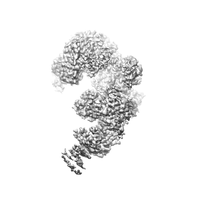
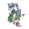
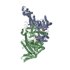
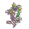
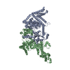
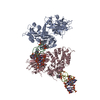
| Title | Cryo-EM structure of the CBF3-core-Ndc10-DBD complex of the budding yeast kinetochore | |||||||||
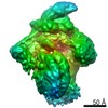
 Map data Map data | CBF3-core-Ndc10-DBD | |||||||||
 Sample Sample |
| |||||||||
| Function / homology |  Function and homology information Function and homology informationRAVE complex / Iron uptake and transport / CBF3 complex / regulation of transcription by galactose / regulation of sulfur amino acid metabolic process / cellular response to methylmercury / vacuolar proton-transporting V-type ATPase complex assembly / FBXL7 down-regulates AURKA during mitotic entry and in early mitosis /  septin ring assembly / mitotic spindle elongation ...RAVE complex / Iron uptake and transport / CBF3 complex / regulation of transcription by galactose / regulation of sulfur amino acid metabolic process / cellular response to methylmercury / vacuolar proton-transporting V-type ATPase complex assembly / FBXL7 down-regulates AURKA during mitotic entry and in early mitosis / septin ring assembly / mitotic spindle elongation / centromeric DNA binding / regulation of exit from mitosis / Antigen processing: Ubiquitination & Proteasome degradation / vacuolar acidification / kinetochore assembly / condensed chromosome, centromeric region / regulation of metabolic process / exit from mitosis / spindle pole body / positive regulation of glucose transmembrane transport / protein neddylation / mitotic intra-S DNA damage checkpoint signaling / silent mating-type cassette heterochromatin formation / mitochondrial fusion / DNA binding, bending / SCF-dependent proteasomal ubiquitin-dependent protein catabolic process / SCF ubiquitin ligase complex / mitotic spindle assembly checkpoint signaling / DNA replication origin binding / regulation of mitotic cell cycle / cullin family protein binding / subtelomeric heterochromatin formation / regulation of protein-containing complex assembly / spindle midzone / endomembrane system / negative regulation of cytoplasmic translation / chromosome segregation / G1/S transition of mitotic cell cycle / spindle / kinetochore / G2/M transition of mitotic cell cycle / mitotic cell cycle / ubiquitin-dependent protein catabolic process / protein-containing complex assembly / chromosome, telomeric region / protein ubiquitination / DNA-binding transcription factor activity, RNA polymerase II-specific / DNA binding / zinc ion binding / identical protein binding / nucleus / cytoplasm septin ring assembly / mitotic spindle elongation ...RAVE complex / Iron uptake and transport / CBF3 complex / regulation of transcription by galactose / regulation of sulfur amino acid metabolic process / cellular response to methylmercury / vacuolar proton-transporting V-type ATPase complex assembly / FBXL7 down-regulates AURKA during mitotic entry and in early mitosis / septin ring assembly / mitotic spindle elongation / centromeric DNA binding / regulation of exit from mitosis / Antigen processing: Ubiquitination & Proteasome degradation / vacuolar acidification / kinetochore assembly / condensed chromosome, centromeric region / regulation of metabolic process / exit from mitosis / spindle pole body / positive regulation of glucose transmembrane transport / protein neddylation / mitotic intra-S DNA damage checkpoint signaling / silent mating-type cassette heterochromatin formation / mitochondrial fusion / DNA binding, bending / SCF-dependent proteasomal ubiquitin-dependent protein catabolic process / SCF ubiquitin ligase complex / mitotic spindle assembly checkpoint signaling / DNA replication origin binding / regulation of mitotic cell cycle / cullin family protein binding / subtelomeric heterochromatin formation / regulation of protein-containing complex assembly / spindle midzone / endomembrane system / negative regulation of cytoplasmic translation / chromosome segregation / G1/S transition of mitotic cell cycle / spindle / kinetochore / G2/M transition of mitotic cell cycle / mitotic cell cycle / ubiquitin-dependent protein catabolic process / protein-containing complex assembly / chromosome, telomeric region / protein ubiquitination / DNA-binding transcription factor activity, RNA polymerase II-specific / DNA binding / zinc ion binding / identical protein binding / nucleus / cytoplasmSimilarity search - Function | |||||||||
| Biological species |  Saccharomyces cerevisiae S288C (yeast) Saccharomyces cerevisiae S288C (yeast) | |||||||||
| Method | single particle reconstruction / cryo EM / Resolution: 3.6 Å | |||||||||
 Authors Authors | Yan K / Zhang Z / Yang J / McLaughlin SH / Barford D | |||||||||
| Funding support |  United Kingdom, 2 items United Kingdom, 2 items
| |||||||||
 Citation Citation | Journal: Nat Struct Mol Biol / Year: 2018 Title: Architecture of the CBF3-centromere complex of the budding yeast kinetochore. Authors: Kaige Yan / Ziguo Zhang / Jing Yang / Stephen H McLaughlin / David Barford / Abstract: Kinetochores are multicomponent complexes responsible for coordinating the attachment of centromeric DNA to mitotic-spindle microtubules. The point centromeres of budding yeast are organized into ...Kinetochores are multicomponent complexes responsible for coordinating the attachment of centromeric DNA to mitotic-spindle microtubules. The point centromeres of budding yeast are organized into three centromeric determining elements (CDEs), and are associated with the centromere-specific nucleosome Cse4. Deposition of Cse4 at CEN loci is dependent on the CBF3 complex that engages CDEIII to direct Cse4 nucleosomes to CDEII. To understand how CBF3 recognizes CDEIII and positions Cse4, we determined a cryo-EM structure of a CBF3-CEN complex. CBF3 interacts with CEN DNA as a head-to-head dimer that includes the whole of CDEIII and immediate 3' regions. Specific CEN-binding of CBF3 is mediated by a Cep3 subunit of one of the CBF3 protomers that forms major groove interactions with the conserved and essential CCG and TGT motifs of CDEIII. We propose a model for a CBF3-Cse4-CEN complex with implications for understanding CBF3-directed deposition of the Cse4 nucleosome at CEN loci. | |||||||||
| History |
|
- Structure visualization
| Movie |
Movie viewer |
|---|---|
| Structure viewer | EM map: SurfViewMolmilJmol/JSmol |
| Supplemental images |
- Downloads & links
-EMDB archive
| Map data | emd_0095.map.gz | 2.8 MB | EMDB map data format | |
|---|---|---|---|---|
| Header (meta data) | emd-0095-v30.xmlemd-0095.xml | 18.4 KB 18.4 KB | Display Display | EMDB header |
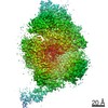
| Images |  emd_0095.png emd_0095.png | 30.7 KB | ||
| Archive directory |  http://ftp.pdbj.org/pub/emdb/structures/EMD-0095ftp://ftp.pdbj.org/pub/emdb/structures/EMD-0095 http://ftp.pdbj.org/pub/emdb/structures/EMD-0095ftp://ftp.pdbj.org/pub/emdb/structures/EMD-0095 | HTTPS FTP |
-Related structure data
| Related structure data | 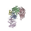 6gypMC 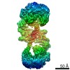 0096C 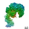 0097C 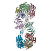 6gysC 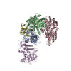 6gyuC M: atomic model generated by this map C: citing same article ( |
|---|---|
| Similar structure data |


-Links
| EMDB pages | EMDB (EBI/PDBe) / EMDataResource |
|---|---|
| Related items in Molecule of the Month |


-Map
| File | Download / File: emd_0095.map.gz / Format: CCP4 / Size: 244.1 MB / Type: IMAGE STORED AS FLOATING POINT NUMBER (4 BYTES) | ||||||||||||||||||||||||||||||||||||||||||||||||||||||||||||||||||||
|---|---|---|---|---|---|---|---|---|---|---|---|---|---|---|---|---|---|---|---|---|---|---|---|---|---|---|---|---|---|---|---|---|---|---|---|---|---|---|---|---|---|---|---|---|---|---|---|---|---|---|---|---|---|---|---|---|---|---|---|---|---|---|---|---|---|---|---|---|---|
| Annotation | CBF3-core-Ndc10-DBD | ||||||||||||||||||||||||||||||||||||||||||||||||||||||||||||||||||||
| Voxel size | X=Y=Z: 1.06 Å | ||||||||||||||||||||||||||||||||||||||||||||||||||||||||||||||||||||
| Density |
| ||||||||||||||||||||||||||||||||||||||||||||||||||||||||||||||||||||
| Symmetry | Space group: 1 | ||||||||||||||||||||||||||||||||||||||||||||||||||||||||||||||||||||
| Details | EMDB XML:
CCP4 map header:
| ||||||||||||||||||||||||||||||||||||||||||||||||||||||||||||||||||||
-Supplemental data
- Sample components
+Entire : CBF3-core-Ndc10-DBD complex
+Supramolecule #1: CBF3-core-Ndc10-DBD complex
 unidentified baculovirus
+Macromolecule #1: Centromere DNA-binding protein complex CBF3 subunit B
+Macromolecule #2: Centromere DNA-binding protein complex CBF3 subunit C
+Macromolecule #3: Centromere DNA-binding protein complex CBF3 subunit B
+Macromolecule #4: Suppressor of kinetochore protein 1
+Macromolecule #5: Centromere DNA-binding protein complex CBF3 subunit A
+Macromolecule #6: METHIONINE

+Macromolecule #7: PHENYLALANINE

+Macromolecule #8: ASPARAGINE
+Macromolecule #9: ARGININE

+Macromolecule #10: THREONINE
+Macromolecule #11: GLUTAMINE

-Experimental details
-Structure determination
| Method | cryo EM |
|---|---|
 Processing Processing | single particle reconstruction |
| Aggregation state | particle |
-Sample preparation
| Concentration | 0.4 mg/mL |
|---|---|
| Buffer | pH: 8 |
| Vitrification | Cryogen name: ETHANE |
- Electron microscopy
| Microscope | FEI TITAN KRIOS |
|---|---|
| Electron beam | Acceleration voltage: 300 kV / Electron source: FIELD EMISSION GUN |
| Electron optics | Illumination mode: FLOOD BEAM / Imaging mode: BRIGHT FIELDBright-field microscopy |
| Image recording | Film or detector model: FEI FALCON III (4k x 4k) / Detector mode: COUNTING / Average electron dose: 27.0 e/Å2 |
| Experimental equipment |  Model: Titan Krios / Image courtesy: FEI Company |
-Image processing
| Startup model | Type of model: OTHER / Details: Initial model was from SIMPLE_PRIME. |
|---|---|
| Initial angle assignment | Type: MAXIMUM LIKELIHOOD |
| Final angle assignment | Type: MAXIMUM LIKELIHOOD |
| Final reconstruction | Resolution.type: BY AUTHOR / Resolution: 3.6 Å / Resolution method: FSC 0.143 CUT-OFF / Number images used: 73894 |
-Atomic model buiding 1
| Refinement | Protocol: OTHER |
|---|---|
| Output model | PDB-6gyp: |
