| タイトル | SAR and cellular potency optimization of novel heme-binding IDO1 inhibitors |
|---|
| ジャーナル・号・ページ | To Be Published |
|---|
| 掲載日 | 2024年3月26日 (構造データの登録日) |
|---|
 著者 著者 | Cren, S. / Lotz, C. / Mac Sweeney, A. / Lange, R. / Kimmerlin, T. |
|---|
 リンク リンク | PubMedで検索 |
|---|
| 手法 | X線回折 |
|---|
| 解像度 | 1.694 - 2.539 Å |
|---|
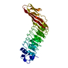
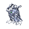
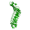
| 構造データ | PDB-9esb:
Holo IDO with a bound inhibitor
手法: X-RAY DIFFRACTION / 解像度: 2.249 Å PDB-9esc:
Holo IDO with a bound inhibitor
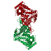
手法: X-RAY DIFFRACTION / 解像度: 1.95 Å PDB-9esd:
Holo TDO with a bound inhibitor
手法: X-RAY DIFFRACTION / 解像度: 2.103 Å PDB-9ese:
Holo IDO with a bound inhibitor
手法: X-RAY DIFFRACTION / 解像度: 2.539 Å PDB-9esf:
Holo IDO with a bound inhibitor
手法: X-RAY DIFFRACTION / 解像度: 2.501 Å PDB-9esg:
Holo IDO with a bound inhibitor
手法: X-RAY DIFFRACTION / 解像度: 2.497 Å PDB-9etv:
Holo IDO with a bound inhibitor
手法: X-RAY DIFFRACTION / 解像度: 2.402 Å PDB-9ew0:
Holo IDO with a bound inhibitor
手法: X-RAY DIFFRACTION / 解像度: 1.8 Å PDB-9f5r:
Holo IDO with a bound inhibitor
手法: X-RAY DIFFRACTION / 解像度: 1.694 Å |
|---|
| 化合物 | PDB-1h6t:
Internalin B: crystal structure of fused N-terminal domains.
PDB-1h6r:
The oxidized state of a redox sensitive variant of green fluorescent protein
ChemComp-5PK:
(1~{R})-1-cyclohexyl-2-[(5~{S})-5~{H}-imidazo[1,5-b]isoindol-5-yl]ethanol
PDB-1h6v:
Mammalian thioredoxin reductase
PDB-1h6u:
Internalin H: crystal structure of fused N-terminal domains.
PDB-1h60:
Structure of Pentaerythritol Tetranitrate Reductase in complex with progesterone
PDB-1h61:
Structure of Pentaerythritol Tetranitrate Reductase in complex with prednisone
PDB-1h93:
ACTIVE MUTANT (S215->C) OF GLUCOSE 6-PHOSPHATE DEHYDROGENASE FROM LEUCONOSTOC MESENTEROIDES
|
|---|
| 由来 |  homo sapiens (ヒト) homo sapiens (ヒト)  drosophila melanogaster (キイロショウジョウバエ) drosophila melanogaster (キイロショウジョウバエ)
|
|---|
 キーワード キーワード | OXIDOREDUCTASE / Inhibitor / heme |
|---|
 ムービー
ムービー コントローラー
 構造ビューア
 万見文献について