[English] 日本語
EMN Papers
- Database of articles cited by 3DEM data entries -

+
Search query

Keywords
Author
Journal
IF

-
Structure paper

TitleDevelopment of a random background to understand ligand optimization.
Journal, issue, pagesNature, Year 2026
Publish dateSep 16, 2026
AuthorsXinyu Xu / Olivier Mailhot / Galen J Correy / Xi-Ping Huang / Joao M Braz / Da Shi / Karthik Srinivasan / Kara Zielinski / Yuliia Holota / Yuliia Kuziv / Christos Iliopoulos-Tsoutsouvas / Nathan D Levinzon / Yagmur U Doruk / Moira M Rachman / Morgan E Diolaiti / Maisie G V Stevens / Fangyu Liu / Katie L Holland / Harald Hübner / Jing Wang / Yujin Wu / Alan Ashworth / Alexandros Makriyannis / Yuqi Zhang / Yurii S Moroz / Peter Gmeiner / Robert Abel / Aashish Manglik / Allan I Basbaum / Bryan L Roth / James S Fraser / Brian K Shoichet /
PubMed AbstractLigand optimization is central to drug discovery, with hundreds of analogues often designed and synthesized between an initial hit and a therapeutic candidate. The efficiency of this process is ...Ligand optimization is central to drug discovery, with hundreds of analogues often designed and synthesized between an initial hit and a therapeutic candidate. The efficiency of this process is unclear, partly because there is no random background for optimization to compare against. Such a random background might emerge from systematic random small substitutions across starting ligands, measuring the likelihood of achieving a substantial improvement in affinity or potency, or other property by any single perturbation. Recent literature has suggested that perhaps 10% of analogues with minor modifications improve upon the potency of a parent by tenfold or more, but this number is clouded by reporting bias, intentional improvement and inter-group variability. To begin to establish a background expectation for ligand optimization, here we systematically modified 18 lead molecules across six targets with single-atom changes; 257 compounds were synthesized. Unexpectedly, 11.3% of these random small perturbation analogues improved potency by tenfold or more. Conversely, they typically had worse in vitro pharmacokinetics. Although it was possible to find analogues where the potency increase compensated for inferior exposure and half-life, resulting in more potent compounds in vivo, overall, a frustrated landscape for ligand optimization is revealed. This study begins to establish a background expectation for ligand potency optimization and offers a simple strategy to do so. It also begins to quantify the challenges confronting the field in moving beyond in vitro potency.
External linksNature / PubMed:42749805
MethodsEM (single particle) / X-ray diffraction
Resolution0.97 - 2.8 Å
Structure data

EMDB-71718, PDB-9pln:
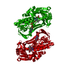
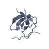
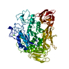
Locally-refined structure of alpha2a adrenergic receptor in complex with Go heterotrimer, scFv16, and N-(5-methylnaphthalen-1-yl)pyridin-4-amine (compound 4905)
Method: EM (single particle) / Resolution: 2.8 Å

EMDB-71719, PDB-9plo:
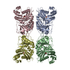
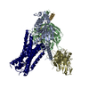
Structure of alpha2a adrenergic receptor in complex with Go heterotrimer, scFv16, and N-(5-methylnaphthalen-1-yl)pyridin-4-amine (compound 4905)
Method: EM (single particle) / Resolution: 2.8 Å

PDB-14ab:
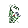
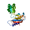
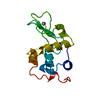
PanDDA analysis group deposition -- Crystal structure of SARS-CoV-2 NSP3 macrodomain in complex with Z1039063794
Method: X-RAY DIFFRACTION / Resolution: 0.98 Å

PDB-14am:
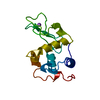
PanDDA analysis group deposition -- Crystal structure of SARS-CoV-2 NSP3 macrodomain in complex with Z8207599372
Method: X-RAY DIFFRACTION / Resolution: 0.99 Å

PDB-14an:
PanDDA analysis group deposition -- Crystal structure of SARS-CoV-2 NSP3 macrodomain in complex with Z8756044539
Method: X-RAY DIFFRACTION / Resolution: 0.98 Å

PDB-14ao:
PanDDA analysis group deposition -- Crystal structure of SARS-CoV-2 NSP3 macrodomain in complex with Z9187344358
Method: X-RAY DIFFRACTION / Resolution: 1.02 Å

PDB-14ap:
PanDDA analysis group deposition -- Crystal structure of SARS-CoV-2 NSP3 macrodomain in complex with Z9187344338
Method: X-RAY DIFFRACTION / Resolution: 1 Å

PDB-7iiw:
PanDDA analysis group deposition -- Crystal structure of SARS-CoV-2 NSP3 macrodomain in complex with Z5398393122
Method: X-RAY DIFFRACTION / Resolution: 0.97 Å

PDB-7iix:
PanDDA analysis group deposition -- Crystal structure of SARS-CoV-2 NSP3 macrodomain in complex with Z7691912366
Method: X-RAY DIFFRACTION / Resolution: 1.02 Å

PDB-7iiy:
PanDDA analysis group deposition -- Crystal structure of SARS-CoV-2 NSP3 macrodomain in complex with Z8598075283
Method: X-RAY DIFFRACTION / Resolution: 1.01 Å

PDB-7iiz:
PanDDA analysis group deposition -- Crystal structure of SARS-CoV-2 NSP3 macrodomain in complex with Z8598075280
Method: X-RAY DIFFRACTION / Resolution: 0.99 Å

PDB-7ij0:
PanDDA analysis group deposition -- Crystal structure of SARS-CoV-2 NSP3 macrodomain in complex with Z8598075284
Method: X-RAY DIFFRACTION / Resolution: 0.98 Å

PDB-7ij1:
PanDDA analysis group deposition -- Crystal structure of SARS-CoV-2 NSP3 macrodomain in complex with Z8598075302
Method: X-RAY DIFFRACTION / Resolution: 1.01 Å

PDB-7ij2:
PanDDA analysis group deposition -- Crystal structure of SARS-CoV-2 NSP3 macrodomain in complex with Z8727396638
Method: X-RAY DIFFRACTION / Resolution: 0.97 Å

PDB-7ij3:
PanDDA analysis group deposition -- Crystal structure of SARS-CoV-2 NSP3 macrodomain in complex with Z8727396582
Method: X-RAY DIFFRACTION / Resolution: 0.97 Å

PDB-7ij4:
PanDDA analysis group deposition -- Crystal structure of SARS-CoV-2 NSP3 macrodomain in complex with Z7692046343
Method: X-RAY DIFFRACTION / Resolution: 1.01 Å

PDB-7ij5:
PanDDA analysis group deposition -- Crystal structure of SARS-CoV-2 NSP3 macrodomain in complex with Z7692056627
Method: X-RAY DIFFRACTION / Resolution: 0.99 Å

PDB-7ij6:
PanDDA analysis group deposition -- Crystal structure of SARS-CoV-2 NSP3 macrodomain in complex with Z7140729870
Method: X-RAY DIFFRACTION / Resolution: 1.02 Å

PDB-7ij7:
PanDDA analysis group deposition -- Crystal structure of SARS-CoV-2 NSP3 macrodomain in complex with Z7691912473
Method: X-RAY DIFFRACTION / Resolution: 1.01 Å

PDB-7ij8:
PanDDA analysis group deposition -- Crystal structure of SARS-CoV-2 NSP3 macrodomain in complex with Z8928826043
Method: X-RAY DIFFRACTION / Resolution: 1 Å

PDB-7ij9:
PanDDA analysis group deposition -- Crystal structure of SARS-CoV-2 NSP3 macrodomain in complex with Z8928826033
Method: X-RAY DIFFRACTION / Resolution: 1 Å

PDB-7ija:
PanDDA analysis group deposition -- Crystal structure of SARS-CoV-2 NSP3 macrodomain in complex with Z8929429249
Method: X-RAY DIFFRACTION / Resolution: 1 Å

PDB-7ijb:
PanDDA analysis group deposition -- Crystal structure of SARS-CoV-2 NSP3 macrodomain in complex with Z8768700676
Method: X-RAY DIFFRACTION / Resolution: 1 Å

PDB-7ijc:
PanDDA analysis group deposition -- Crystal structure of SARS-CoV-2 NSP3 macrodomain in complex with Z8929428675
Method: X-RAY DIFFRACTION / Resolution: 1 Å

PDB-7ijd:
PanDDA analysis group deposition -- Crystal structure of SARS-CoV-2 NSP3 macrodomain in complex with Z7692056404
Method: X-RAY DIFFRACTION / Resolution: 1 Å

PDB-7ije:
PanDDA analysis group deposition -- Crystal structure of SARS-CoV-2 NSP3 macrodomain in complex with Z8990523169
Method: X-RAY DIFFRACTION / Resolution: 1 Å

PDB-7ijf:
PanDDA analysis group deposition -- Crystal structure of SARS-CoV-2 NSP3 macrodomain in complex with Z8727401304
Method: X-RAY DIFFRACTION / Resolution: 1 Å

PDB-7ijg:
PanDDA analysis group deposition -- Crystal structure of SARS-CoV-2 NSP3 macrodomain in complex with Z8990527720
Method: X-RAY DIFFRACTION / Resolution: 1.02 Å

PDB-7ijh:
PanDDA analysis group deposition -- Crystal structure of SARS-CoV-2 NSP3 macrodomain in complex with Z8990523176
Method: X-RAY DIFFRACTION / Resolution: 1 Å

PDB-7iji:
PanDDA analysis group deposition -- Crystal structure of SARS-CoV-2 NSP3 macrodomain in complex with Z8990523172
Method: X-RAY DIFFRACTION / Resolution: 1.02 Å

PDB-7ijj:
PanDDA analysis group deposition -- Crystal structure of SARS-CoV-2 NSP3 macrodomain in complex with Z8990919037
Method: X-RAY DIFFRACTION / Resolution: 0.98 Å

PDB-7ijk:
PanDDA analysis group deposition -- Crystal structure of SARS-CoV-2 NSP3 macrodomain in complex with Z8990919048
Method: X-RAY DIFFRACTION / Resolution: 0.98 Å

PDB-7ijl:
PanDDA analysis group deposition -- Crystal structure of SARS-CoV-2 NSP3 macrodomain in complex with Z8990523194
Method: X-RAY DIFFRACTION / Resolution: 1.01 Å

PDB-7ijm:
PanDDA analysis group deposition -- Crystal structure of SARS-CoV-2 NSP3 macrodomain in complex with Z7534253453
Method: X-RAY DIFFRACTION / Resolution: 1.01 Å

Chemicals

PDB-1dbs:
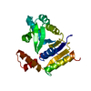
MECHANISTIC IMPLICATIONS AND FAMILY RELATIONSHIPS FROM THE STRUCTURE OF DETHIOBIOTIN SYNTHETASE

ChemComp-HOH:
WATER

PDB-1dbl:
Unknown entry

PDB-1dbm:
MOLECULAR BASIS OF CROSS-REACTIVITY AND THE LIMITS OF ANTIBODY-ANTIGEN COMPLEMENTARITY

ChemComp-DMS:
DIMETHYL SULFOXIDE / DMSO, precipitant*YM

PDB-1dbn:
MAACKIA AMURENSIS LEUKOAGGLUTININ (LECTIN) WITH SIALYLLACTOSE

PDB-1dbe:
Unknown entry

PDB-1dbd:
E2 DNA-BINDING DOMAIN FROM PAPILLOMAVIRUS BPV-1

ChemComp-RLU:
(3R)-3-(4-bromophenyl)-3-[(7H-pyrrolo[2,3-d]pyrimidine-4-carbonyl)amino]propanoic acid

PDB-1cjb:
MALARIAL PURINE PHOSPHORIBOSYLTRANSFERASE

PDB-1cjc:
STRUCTURE OF ADRENODOXIN REDUCTASE OF MITOCHONDRIAL P450 SYSTEMS

PDB-1cjd:
THE BACTERIOPHAGE PRD1 COAT PROTEIN, P3, IS STRUCTURALLY SIMILAR TO HUMAN ADENOVIRUS HEXON

PDB-1cje:
ADRENODOXIN FROM BOVINE

PDB-1cjf:
PROFILIN BINDS PROLINE-RICH LIGANDS IN TWO DISTINCT AMIDE BACKBONE ORIENTATIONS

PDB-1cjr:
X-RAY CRYSTALLOGRAPHIC STUDIES OF DENATURATION IN RIBONUCLEASE S

PDB-1cjs:
CRYSTAL STRUCTURE OF RIBOSOMAL PROTEIN L1 FROM METHANOCOCCUS JANNASCHII

PDB-1cjt:
COMPLEX OF GS-ALPHA WITH THE CATALYTIC DOMAINS OF MAMMALIAN ADENYLYL CYCLASE: COMPLEX WITH BETA-L-2',3'-DIDEOXYATP, MN, AND MG

PDB-1cjv:
COMPLEX OF GS-ALPHA WITH THE CATALYTIC DOMAINS OF MAMMALIAN ADENYLYL CYCLASE: COMPLEX WITH BETA-L-2',3'-DIDEOXYATP, MG, AND ZN

PDB-1cjw:
SEROTONIN N-ACETYLTRANSFERASE COMPLEXED WITH A BISUBSTRATE ANALOG

PDB-1cjx:
CRYSTAL STRUCTURE OF PSEUDOMONAS FLUORESCENS HPPD

PDB-1cjy:
HUMAN CYTOSOLIC PHOSPHOLIPASE A2


PDB Unreleased entry

PDB-1cjz: COMPLEX OF GS-ALPHA WITH THE CATALYTIC DOMAINS OF MAMMALIAN ADENYLYL CYCLASE: COMPLEX WITH BETA-L-2'',3''-DIDEOXYATP, MN, AND MG
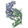
PDB-1cj0:
CRYSTAL STRUCTURE OF RABBIT CYTOSOLIC SERINE HYDROXYMETHYLTRANSFERASE AT 2.8 ANGSTROM RESOLUTION

PDB-1cj1:
GROWTH FACTOR RECEPTOR BINDING PROTEIN SH2 DOMAIN (HUMAN) COMPLEXED WITH A PHOSPHOTYROSYL DERIVATIVE

PDB-1cj3:
MUTANT TYR38GLU OF PARA-HYDROXYBENZOATE HYDROXYLASE

PDB-1akg:
ALPHA-CONOTOXIN PNIB FROM CONUS PENNACEUS

PDB-1cj4:
MUTANT Q34T OF PARA-HYDROXYBENZOATE HYDROXYLASE

PDB-1cj5:
BOVINE BETA-LACTOGLOBULIN A

PDB-1cj6:
T11A MUTANT HUMAN LYSOZYME

PDB-1cj7:
T11V MUTANT HUMAN LYSOZYME

PDB-1cj8:
T40A MUTANT HUMAN LYSOZYME

PDB-1cka:
STRUCTURAL BASIS FOR THE SPECIFIC INTERACTION OF LYSINE-CONTAINING PROLINE-RICH PEPTIDES WITH THE N-TERMINAL SH3 DOMAIN OF C-CRK

PDB-1ckb:
STRUCTURAL BASIS FOR THE SPECIFIC INTERACTION OF LYSINE-CONTAINING PROLINE-RICH PEPTIDES WITH THE N-TERMINAL SH3 DOMAIN OF C-CRK

PDB-1ckc:
T43A MUTANT HUMAN LYSOZYME

PDB-1ckd:
T43V MUTANT HUMAN LYSOZYME

PDB-1cke:
CMP KINASE FROM ESCHERICHIA COLI FREE ENZYME STRUCTURE

PDB-1ckf:
T52A MUTANT HUMAN LYSOZYME

PDB-1ckh:
T70V MUTANT HUMAN LYSOZYME

PDB-1cki:
RECOMBINANT CASEIN KINASE I DELTA TRUNCATION MUTANT CONTAINING RESIDUES 1-317

PDB-1ckj:
CASEIN KINASE I DELTA TRUNCATION MUTANT CONTAINING RESIDUES 1-317 COMPLEX WITH BOUND TUNGSTATE

PDB-1ckk:
CALMODULIN/RAT CA2+/CALMODULIN DEPENDENT PROTEIN KINASE FRAGMENT

PDB-1ckl:
N-TERMINAL TWO DOMAINS OF HUMAN CD46 (MEMBRANE COFACTOR PROTEIN, MCP)

PDB-1ciu:
THERMOSTABLE CGTASE FROM THERMOANAEROBACTERIUM THERMOSULFURIGENES EM1 AT PH 8.0.

Source
  • homo sapiens (human)
  • mus musculus (house mouse)
  • severe acute respiratory syndrome coronavirus 2
KeywordsVIRAL PROTEIN / Macrodomain / ADP-ribose / SARS-CoV-2 / fragment-based drug discovery / MEMBRANE PROTEIN / Receptor / Drug / Complex / GPCR / G-protein / scFv16

+
About EMN Papers

-
News

-
Feb 9, 2022. New format data for meta-information of EMDB entries

New format data for meta-information of EMDB entries

  • Version 3 of the EMDB header file is now the official format.
  • The previous official version 1.9 will be removed from the archive.

Related info.:EMDB header

External links:wwPDB to switch to version 3 of the EMDB data model

-
Oct 5, 2021. Nobel Prize for mechanically activated and temperature-gated ion channels

Nobel Prize for mechanically activated and temperature-gated ion channels

  • The Nobel Prize in Physiology or Medicine 2021 was awarded jointly to David Julius and Ardem Patapoutian "for their discoveries of receptors for temperature and touch."
  • EM Navigator can help to find cryo-EM structure data by both pioneers.

External links:The Nobel Prize in Physiology or Medicine 2021 - NobelPrize.org / Structure data by Ardem Patapoutian / Structure data by David Julius

+
Aug 12, 2020. Covid-19 info

Covid-19 info

URL: https://pdbj.org/emnavi/covid19.php

New page: Covid-19 featured information page in EM Navigator.

Related info.:Covid-19 info / Mar 5, 2020. Novel coronavirus structure data

+
Mar 5, 2020. Novel coronavirus structure data

Novel coronavirus structure data

Related info.:Yorodumi Speices / Aug 12, 2020. Covid-19 info

External links:COVID-19 featured content - PDBj / Molecule of the Month (242):Coronavirus Proteases

+
Jul 5, 2019. Downlodablable text data

Downlodablable text data

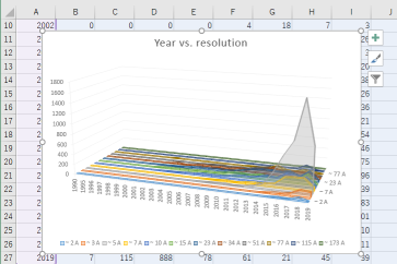
Some data of EM Navigator services can be downloaded as text file. Software such as Excel can load the data files.

PageDataFormat
EMN Searchsearch resultCSV, TSV, or JSON
EMN statisticsdata tableCSV or TSV

Related info.:EMN Search / EMN Statistics

-
EMN Papers

Database of articles cited by 3DEM data entries

  • Database of articles cited by 3DEM data entries in EMDB and PDB
  • Using PubMed data

Related info.:EMDB / PDB / Q: What are the data sources of EM Navigator? / EM Navigator / Yorodumi Papers / Changes in new EM Navigator and Yorodumi

Read more