 ムービー
ムービー コントローラー
 構造ビューア
 EMN検索について


-検索条件
-検索結果
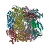
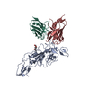
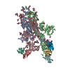
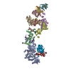
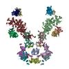
検索 (著者・登録者: j. & e. & johnson)の結果51件中、1から50件目までを表示しています

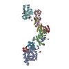
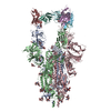
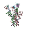
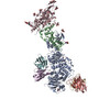
PDB-8fai: 
Cryo-EM structure of the Agrobacterium T-pilus
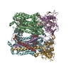
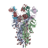
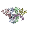
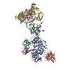
PDB-8u7i:
Structure of the phage immune evasion protein Gad1 bound to the Gabija GajAB complex
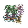
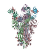
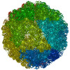
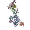
PDB-8asi:
Four subunit cytochrome b-c1 complex from Rhodobacter sphaeroides in native nanodiscs - consensus refinement in the b-b conformation
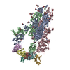
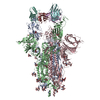
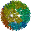
PDB-8asj:
Four subunit cytochrome b-c1 complex from Rhodobacter sphaeroides in native nanodiscs - focussed refinement in the b-c conformation

PDB-8a3c:
Nudaurelia capensis omega virus maturation intermediate captured at pH5.9 (insect cell expressed VLPs)

PDB-8a41:
Nudaurelia capensis omega virus procapsid at pH7.6 (insect cell expressed VLPs)

PDB-8a6j:
Nudaurelia capensis omega virus maturation intermediate captured at pH6.25 (insect cell expressed VLPs)

PDB-8aay:
Nudaurelia capensis omega virus maturation intermediate captured at pH5.6 (insect cell expressed VLPs): small class from symmetry expansion

PDB-8ac6:
Nudaurelia capensis omega virus maturation intermediate captured at pH5.6 (insect cell expressed VLPs): medium class from symmetry expansion

PDB-8ach:
Nudaurelia capensis omega virus maturation intermediate captured at pH5.6 (insect cell expressed VLPs): large class from symmetry expansion
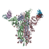
PDB-7zr7:
OMI-42 FAB IN COMPLEX WITH SARS-COV-2 BETA SPIKE GLYCOPROTEIN
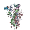
PDB-7zr8:
OMI-38 FAB IN COMPLEX WITH SARS-COV-2 BETA SPIKE RBD (local refinement)

PDB-7zr9:
OMI-2 FAB IN COMPLEX WITH SARS-COV-2 BETA SPIKE GLYCOPROTEIN
PDB-7zrc:
OMI-38 FAB IN COMPLEX WITH SARS-COV-2 BETA SPIKE
PDB-7t4q:
CryoEM structure of the HCMV Pentamer gH/gL/UL128/UL130/UL131A in complex with neutralizing fabs 2C12, 7I13 and 13H11
PDB-7t4r:
CryoEM structure of the HCMV Pentamer gH/gL/UL128/UL130/UL131A in complex with THBD and neutralizing fabs MSL-109 and 13H11
PDB-7t4s:
CryoEM structure of the HCMV Pentamer gH/gL/UL128/UL130/UL131A in complex with NRP2 and neutralizing fabs 8I21 and 13H11
PDB-7q6e:
Beta049 fab in complex with SARS-CoV2 beta-Spike glycoprotein, The Beta mAb response underscores the antigenic distance to other SARS-CoV-2 variants
PDB-7q9f:
Beta-50 fab in complex with SARS-CoV-2 beta-Spike glycoprotein
PDB-7q9g:
COVOX-222 fab in complex with SARS-CoV-2 beta-Spike glycoprotein
PDB-7q9i:
Beta-43 fab in complex with SARS-CoV-2 beta-Spike glycoprotein
PDB-7q9j:
Beta-26 fab in complex with SARS-CoV-2 beta-Spike glycoprotein
PDB-7q9k:
Beta-32 fab in complex with SARS-CoV-2 beta-Spike glycoprotein
PDB-7q9m:
Beta-53 fab in complex with SARS-CoV-2 beta-Spike glycoprotein
PDB-7q9p:
Beta-06 fab in complex with SARS-CoV-2 beta-Spike glycoprotein
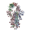
PDB-7kw7:
Atomic cryoEM structure of Hsp90-Hsp70-Hop-GR
PDB-7anm:
Nudaurelia capensis omega virus capsid: virus-like particles expressed in Nicotiana benthamiana
PDB-7ata:
Nudaurelia capensis omega virus procapsid: virus-like particles expressed in Nicotiana benthamiana

PDB-7akv:
The cryo-EM structure of the Vag8-C1 inhibitor complex
PDB-7lbe:
CryoEM structure of the HCMV Trimer gHgLgO in complex with neutralizing fabs 13H11 and MSL-109
PDB-7lbf:
CryoEM structure of the HCMV Trimer gHgLgO in complex with human Platelet-derived growth factor receptor alpha and neutralizing fabs 13H11 and MSL-109
PDB-7lbg:
CryoEM structure of the HCMV Trimer gHgLgO in complex with human Transforming growth factor beta receptor type 3 and neutralizing fabs 13H11 and MSL-109
PDB-6oig:
Subunit joining exposes nascent pre-40S rRNA for processing and quality control

PDB-6wdr:
Subunit joining exposes nascent pre-40S rRNA for processing and quality control
PDB-6scn:
33mer structure of the Salmonella flagella MS-ring protein FliF
PDB-6sd1:
Structure of the RBM3/collar region of the Salmonella flagella MS-ring protein FliF with 33-fold symmetry applied
PDB-6sd2:
Structure of the RBM2inner region of the Salmonella flagella MS-ring protein FliF with 21-fold symmetry applied.
PDB-6sd3:
34mer structure of the Salmonella flagella MS-ring protein FliF
PDB-6sd4:
Structure of the RBM3/collar region of the Salmonella flagella MS-ring protein FliF with 34-fold symmetry applied
PDB-6sd5:
Structure of the RBM2 inner ring of Salmonella flagella MS-ring protein FliF with 22-fold symmetry applied
PDB-6tre:
Structure of the RBM3/collar region of the Salmonella flagella MS-ring protein FliF with 32-fold symmetry applied

PDB-6rqf:
3.6 Angstrom cryo-EM structure of the dimeric cytochrome b6f complex from Spinacia oleracea with natively bound thylakoid lipids and plastoquinone molecules
PDB-4znn:
MicroED structure of the segment, GVVHGVTTVA, from the A53T familial mutant of Parkinson's disease protein, alpha-synuclein residues 47-56
PDB-4ril:
Structure of the amyloid forming segment, GAVVTGVTAVA, from the NAC domain of Parkinson's disease protein alpha-synuclein, residues 68-78, determined by electron diffraction

PDB-3j31:
Life in the extremes: atomic structure of Sulfolobus Turreted Icosahedral Virus
PDB-3iyh:
P22 procapsid coat protein structures reveal a novel mechanism for capsid maturation: Stability without auxiliary proteins or chemical cross-links
PDB-3iyi:
P22 expanded head coat protein structures reveal a novel mechanism for capsid maturation: Stability without auxiliary proteins or chemical cross-links

PDB-3ddx:
HK97 bacteriophage capsid Expansion Intermediate-II model

PDB-2v6l:
Molecular Model of a Type III Secretion System Needle

PDB-2fte:
Bacteriophage HK97 Expansion Intermediate IV
ページ:
 wwPDBはEMDBデータモデルのバージョン3へ移行します
