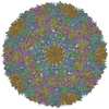
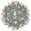
 Movie
Movie Controller

[English] 日本語
 Yorodumi
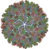
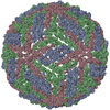
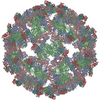
- PDB-6b0x: Capsid protein and C-terminal part of scaffolding protein in the ... -
+ Open data

- Basic information
| Entry | Database: PDB / ID: 6b0x | ||||||
|---|---|---|---|---|---|---|---|
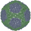
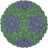
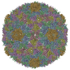
| Title | Capsid protein and C-terminal part of scaffolding protein in the Staphylococcus aureus phage 80alpha procapsid | ||||||
 Components Components |
| ||||||
 Keywords Keywords |  VIRUS / major capsid protein / HK97-like fold / scaffolding protein / procapsid VIRUS / major capsid protein / HK97-like fold / scaffolding protein / procapsid | ||||||
| Function / homology | Protein of unknown function DUF4355 / Domain of unknown function (DUF4355) / viral scaffold / Phage capsid / Phage capsid family / Scaffold protein / Major capsid protein Function and homology information Function and homology information | ||||||
| Biological species |  Staphylococcus phage 80alpha (virus) Staphylococcus phage 80alpha (virus) | ||||||
| Method | ELECTRON MICROSCOPY / single particle reconstruction / cryo EM / Resolution: 3.8 Å | ||||||
 Authors Authors | Kizziah, J.L. / Dearborn, A.D. / Dokland, T. | ||||||
| Funding support |  United States, 1items United States, 1items
| ||||||
 Citation Citation | Journal: Elife / Year: 2017 Title: Competing scaffolding proteins determine capsid size during mobilization of pathogenicity islands. Authors: Altaira D Dearborn / Erin A Wall / James L Kizziah / Laura Klenow / Laura K Parker / Keith A Manning / Michael S Spilman / John M Spear / Gail E Christie / Terje Dokland / Abstract: pathogenicity islands (SaPIs), such as SaPI1, exploit specific helper bacteriophages, like 80α, for their high frequency mobilization, a process termed 'molecular piracy'. SaPI1 redirects the ... pathogenicity islands (SaPIs), such as SaPI1, exploit specific helper bacteriophages, like 80α, for their high frequency mobilization, a process termed 'molecular piracy'. SaPI1 redirects the helper's assembly pathway to form small capsids that can only accommodate the smaller SaPI1 genome, but not a complete phage genome. SaPI1 encodes two proteins, CpmA and CpmB, that are responsible for this size redirection. We have determined the structures of the 80α and SaPI1 procapsids to near-atomic resolution by cryo-electron microscopy, and show that CpmB competes with the 80α scaffolding protein (SP) for a binding site on the capsid protein (CP), and works by altering the angle between capsomers. We probed these interactions genetically and identified second-site suppressors of lethal mutations in SP. Our structures show, for the first time, the detailed interactions between SP and CP in a bacteriophage, providing unique insights into macromolecular assembly processes. | ||||||
| History |
|
- Structure visualization
| Movie |
Movie viewer |
|---|---|
| Structure viewer | Molecule: MolmilJmol/JSmol |
- Downloads & links
-Download
| PDBx/mmCIF format | 6b0x.cif.gz | 380.8 KB | Display | PDBx/mmCIF format |
|---|---|---|---|---|
| PDB format | pdb6b0x.ent.gz | 293.6 KB | Display | PDB format |
| PDBx/mmJSON format | 6b0x.json.gz | Tree view | PDBx/mmJSON format | |
| Others |  Other downloads Other downloads |
-Validation report
| Arichive directory | https://data.pdbj.org/pub/pdb/validation_reports/b0/6b0xftp://data.pdbj.org/pub/pdb/validation_reports/b0/6b0x | HTTPS FTP |
|---|
-Related structure data
| Related structure data |  7030MC 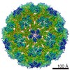 7035C  6b23C M: map data used to model this data C: citing same article ( |
|---|---|
| Similar structure data |
-Links
 PDBj
PDBj

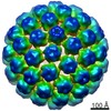
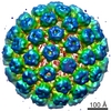
- Assembly
| Deposited unit | 
|
|---|---|
| 1 | x 60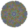
|
| 2 |
|
| 3 | x 5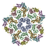
|
| 4 | x 6
|
| 5 | 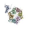
|
| Symmetry | Point symmetry: (Schoenflies symbol: I (icosahedral)) |
-Components
| #1: Protein | Mass: 36846.883 Da / Num. of mol.: 7 Source method: isolated from a genetically manipulated source Source: (gene. exp.) Staphylococcus phage 80alpha (virus) / Gene: orf47 / Cell line (production host): RN4220 / Production host:   Staphylococcus aureus (bacteria) / References: UniProt: A4ZFB3 Staphylococcus aureus (bacteria) / References: UniProt: A4ZFB3#2: Protein | Mass: 23410.941 Da / Num. of mol.: 7 Source method: isolated from a genetically manipulated source Source: (gene. exp.) Staphylococcus phage 80alpha (virus) / Cell line (production host): RN4220 / Production host: Staphylococcus aureus (bacteria) / References: UniProt: A4ZFB2 |
|---|
-Experimental details
-Experiment
| Experiment | Method: ELECTRON MICROSCOPY |
|---|---|
| EM experiment | Aggregation state: PARTICLE / 3D reconstruction method: single particle reconstruction |
- Sample preparation
| Component | Name: Staphylococcus phage 80alpha / Type: VIRUS Details: Lysogenic 80alpha with small terminase gene deletion Entity ID: all / Source: RECOMBINANT |
|---|---|
| Molecular weight | Value: 25.27 MDa / Experimental value: NO |
| Source (natural) | Organism: Staphylococcus phage 80alpha (virus) |
| Source (recombinant) | Organism: Staphylococcus aureus (bacteria) / Strain: RN4220 |
| Details of virus | Empty: YES / Enveloped: NO / Isolate: SPECIES / Type: VIRION |
| Natural host | Organism: Staphylococcus aureus |
| Virus shell | Name: ProcapsidCapsid / Diameter: 546 nm / Triangulation number (T number): 7 |
| Buffer solution | pH: 7.8 |
| Specimen | Embedding applied: NO / Shadowing applied: NO / Staining applied: NO / Vitrification applied: YES |
| Vitrification | Cryogen name: ETHANE |
- Electron microscopy imaging
| Experimental equipment |  Model: Titan Krios / Image courtesy: FEI Company |
|---|---|
| Microscopy | Model: FEI TITAN KRIOS |
| Electron gun | Electron source: FIELD EMISSION GUN / Accelerating voltage: 300 kV / Illumination mode: FLOOD BEAM |
| Electron lens | Mode: BRIGHT FIELDBright-field microscopy |
| Image recording | Electron dose: 40 e/Å2 / Film or detector model: DIRECT ELECTRON DE-20 (5k x 3k) |
- Processing
| Software | Name: REFMAC / Version: 5.8.0088 / Classification: refinement | ||||||||||||||||||||||||||||||||||||||||||||||||||||||||||||||||||||||||||||||||||||||||||||||||||||||||||
|---|---|---|---|---|---|---|---|---|---|---|---|---|---|---|---|---|---|---|---|---|---|---|---|---|---|---|---|---|---|---|---|---|---|---|---|---|---|---|---|---|---|---|---|---|---|---|---|---|---|---|---|---|---|---|---|---|---|---|---|---|---|---|---|---|---|---|---|---|---|---|---|---|---|---|---|---|---|---|---|---|---|---|---|---|---|---|---|---|---|---|---|---|---|---|---|---|---|---|---|---|---|---|---|---|---|---|---|
| CTF correction | Type: PHASE FLIPPING ONLY | ||||||||||||||||||||||||||||||||||||||||||||||||||||||||||||||||||||||||||||||||||||||||||||||||||||||||||
| Symmetry | Point symmetry: I (icosahedral) | ||||||||||||||||||||||||||||||||||||||||||||||||||||||||||||||||||||||||||||||||||||||||||||||||||||||||||
| 3D reconstruction | Resolution: 3.8 Å / Resolution method: FSC 0.143 CUT-OFF / Num. of particles: 10557 / Symmetry type: POINT | ||||||||||||||||||||||||||||||||||||||||||||||||||||||||||||||||||||||||||||||||||||||||||||||||||||||||||
| Refinement | Resolution: 3.8→3.8 Å / Cor.coef. Fo:Fc: 0.88 / SU B: 67.147 / SU ML: 0.83 / ESU R: 0.904 Stereochemistry target values: MAXIMUM LIKELIHOOD WITH PHASES Details: HYDROGENS HAVE BEEN ADDED IN THE RIDING POSITIONS
| ||||||||||||||||||||||||||||||||||||||||||||||||||||||||||||||||||||||||||||||||||||||||||||||||||||||||||
| Solvent computation | Ion probe radii: 0.8 Å / Shrinkage radii: 0.8 Å / VDW probe radii: 1.2 Å / Solvent model: MASK | ||||||||||||||||||||||||||||||||||||||||||||||||||||||||||||||||||||||||||||||||||||||||||||||||||||||||||
| Displacement parameters | Biso mean: 59.505 Å2
| ||||||||||||||||||||||||||||||||||||||||||||||||||||||||||||||||||||||||||||||||||||||||||||||||||||||||||
| Refine LS restraints |
|