 Movie
Movie Controller

[English] 日本語
 Yorodumi
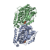
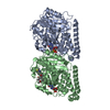
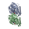
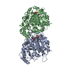
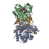
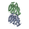
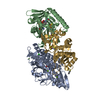
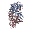
- PDB-5w3j: Yeast microtubule stabilized with Taxol assembled from mutated tubulin -
+ Open data

- Basic information
| Entry | Database: PDB / ID: 5w3j | ||||||||||||||||||||||||
|---|---|---|---|---|---|---|---|---|---|---|---|---|---|---|---|---|---|---|---|---|---|---|---|---|---|
| Title | Yeast microtubule stabilized with Taxol assembled from mutated tubulin | ||||||||||||||||||||||||
 Components Components |
| ||||||||||||||||||||||||
 Keywords Keywords |  HYDROLASE / Cytoskeleton / tubulin HYDROLASE / Cytoskeleton / tubulin | ||||||||||||||||||||||||
| Function / homology |  Function and homology information Function and homology informationnuclear migration by microtubule mediated pushing forces / Cilium Assembly / nuclear division / Sealing of the nuclear envelope (NE) by ESCRT-III / mitotic spindle elongation / nuclear migration along microtubule / homologous chromosome segregation / Platelet degranulation / positive regulation of intracellular protein transport / tubulin complex ...nuclear migration by microtubule mediated pushing forces / Cilium Assembly / nuclear division / Sealing of the nuclear envelope (NE) by ESCRT-III / mitotic spindle elongation / nuclear migration along microtubule / homologous chromosome segregation / Platelet degranulation / positive regulation of intracellular protein transport / tubulin complex / mitotic sister chromatid segregation / mitotic spindle assembly / microtubule-based process / cytoplasmic microtubule organization / cytoskeleton organization / Neutrophil degranulation / nuclear periphery / Hydrolases; Acting on acid anhydrides; Acting on GTP to facilitate cellular and subcellular movement / structural constituent of cytoskeleton / spindle / microtubule cytoskeleton organization / mitotic cell cycle / microtubule / hydrolase activity / response to antibiotic / GTPase activity / GTP binding / metal ion binding / nucleus / cytoplasmSimilarity search - Function | ||||||||||||||||||||||||
| Biological species |  Saccharomyces cerevisiae (brewer's yeast) Saccharomyces cerevisiae (brewer's yeast) | ||||||||||||||||||||||||
| Method | ELECTRON MICROSCOPY / helical reconstruction / cryo EM / Resolution: 4 Å | ||||||||||||||||||||||||
 Authors Authors | Howes, S.C. / Geyer, E.A. / LaFrance, B. / Zhang, R. / Kellogg, E.H. / Westermann, S. / Rice, L.M. / Nogales, E. | ||||||||||||||||||||||||
| Funding support |  United States, 7items United States, 7items
| ||||||||||||||||||||||||
 Citation Citation | Journal: J Cell Biol / Year: 2017 Title: Structural differences between yeast and mammalian microtubules revealed by cryo-EM. Authors: Stuart C Howes / Elisabeth A Geyer / Benjamin LaFrance / Rui Zhang / Elizabeth H Kellogg / Stefan Westermann / Luke M Rice / Eva Nogales /  Abstract: Microtubules are polymers of αβ-tubulin heterodimers essential for all eukaryotes. Despite sequence conservation, there are significant structural differences between microtubules assembled in ...Microtubules are polymers of αβ-tubulin heterodimers essential for all eukaryotes. Despite sequence conservation, there are significant structural differences between microtubules assembled in vitro from mammalian or budding yeast tubulin. Yeast MTs were not observed to undergo compaction at the interdimer interface as seen for mammalian microtubules upon GTP hydrolysis. Lack of compaction might reflect slower GTP hydrolysis or a different degree of allosteric coupling in the lattice. The microtubule plus end-tracking protein Bim1 binds yeast microtubules both between αβ-tubulin heterodimers, as seen for other organisms, and within tubulin dimers, but binds mammalian tubulin only at interdimer contacts. At the concentrations used in cryo-electron microscopy, Bim1 causes the compaction of yeast microtubules and induces their rapid disassembly. Our studies demonstrate structural differences between yeast and mammalian microtubules that likely underlie their differing polymerization dynamics. These differences may reflect adaptations to the demands of different cell size or range of physiological growth temperatures. | ||||||||||||||||||||||||
| History |
|
- Structure visualization
| Movie |
Movie viewer |
|---|---|
| Structure viewer | Molecule: MolmilJmol/JSmol |
- Downloads & links
-Download
| PDBx/mmCIF format | 5w3j.cif.gz | 188.7 KB | Display | PDBx/mmCIF format |
|---|---|---|---|---|
| PDB format | pdb5w3j.ent.gz | 145.9 KB | Display | PDB format |
| PDBx/mmJSON format | 5w3j.json.gz | Tree view | PDBx/mmJSON format | |
| Others |  Other downloads Other downloads |
-Validation report
| Arichive directory | https://data.pdbj.org/pub/pdb/validation_reports/w3/5w3jftp://data.pdbj.org/pub/pdb/validation_reports/w3/5w3j | HTTPS FTP |
|---|
-Related structure data
| Related structure data |  8757MC 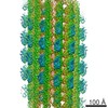 8755C 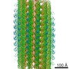 8756C 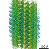 8758C 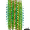 8759C  5w3fC  5w3hC M: map data used to model this data C: citing same article ( |
|---|---|
| Similar structure data |


-Links
 PDBj
PDBj


- Assembly
| Deposited unit | 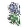
|
|---|---|
| 1 |
|
-Components
-Protein , 2 types, 2 molecules AB
| #1: Protein | Mass: 49853.867 Da / Num. of mol.: 1 Source method: isolated from a genetically manipulated source Source: (gene. exp.) Saccharomyces cerevisiae (strain ATCC 204508 / S288c) (yeast)Production host: Saccharomyces cerevisiae (strain ATCC 204508 / S288c) (yeast)References: UniProt: P09733 |
|---|---|
| #2: Protein | Mass: 51089.668 Da / Num. of mol.: 1 / Mutation: A19K, T23V, G26D, N227H, Y270F Source method: isolated from a genetically manipulated source Source: (gene. exp.) Saccharomyces cerevisiae (strain ATCC 204508 / S288c) (yeast)Production host: Saccharomyces cerevisiae (strain ATCC 204508 / S288c) (yeast)References: UniProt: P02557 |
-Non-polymers , 4 types, 4 molecules 


| #3: Chemical | ChemComp-GTP / Guanosine triphosphate Mass: 523.180 Da / Num. of mol.: 1 / Source method: obtained synthetically / Formula: C10H16N5O14P3 / Comment: GTP, energy-carrying molecule*YM Mass: 523.180 Da / Num. of mol.: 1 / Source method: obtained synthetically / Formula: C10H16N5O14P3 / Comment: GTP, energy-carrying molecule*YM |
|---|---|
| #4: Chemical | ChemComp-MG /  Mass: 24.305 Da / Num. of mol.: 1 / Source method: obtained synthetically / Formula: Mg Mass: 24.305 Da / Num. of mol.: 1 / Source method: obtained synthetically / Formula: Mg |
| #5: Chemical | ChemComp-GDP / Guanosine diphosphate Type: RNA linking / Mass: 443.201 Da / Num. of mol.: 1 / Source method: obtained synthetically / Formula: C10H15N5O11P2 / Comment: GDP, energy-carrying molecule*YM Type: RNA linking / Mass: 443.201 Da / Num. of mol.: 1 / Source method: obtained synthetically / Formula: C10H15N5O11P2 / Comment: GDP, energy-carrying molecule*YM |
| #6: Chemical | ChemComp-TA1 / Paclitaxel Mass: 853.906 Da / Num. of mol.: 1 / Source method: obtained synthetically / Formula: C47H51NO14 / Comment: medication, chemotherapy*YM Mass: 853.906 Da / Num. of mol.: 1 / Source method: obtained synthetically / Formula: C47H51NO14 / Comment: medication, chemotherapy*YM |
-Experimental details
-Experiment
| Experiment | Method: ELECTRON MICROSCOPY |
|---|---|
| EM experiment | Aggregation state: HELICAL ARRAY / 3D reconstruction method: helical reconstruction |
- Sample preparation
| Component | Name: Yeast microtubule stabilized with taxol assembled from mutated tubulin Type: COMPLEX / Entity ID: #1-#2 / Source: NATURAL |
|---|---|
| Molecular weight | Experimental value: NO |
| Source (natural) | Organism: Saccharomyces cerevisiae S288c (yeast) |
| Buffer solution | pH: 6.9 |
| Specimen | Embedding applied: NO / Shadowing applied: NO / Staining applied: NO / Vitrification applied: YES |
| Vitrification | Instrument: FEI VITROBOT MARK IV / Cryogen name: ETHANE / Humidity: 100 % / Chamber temperature: 303 K |
- Electron microscopy imaging
| Microscopy | Model: FEI TITAN |
|---|---|
| Electron gun | Electron source: FIELD EMISSION GUN / Accelerating voltage: 300 kV / Illumination mode: FLOOD BEAM |
| Electron lens | Mode: BRIGHT FIELDBright-field microscopy |
| Specimen holder | Cryogen: NITROGEN Specimen holder model: GATAN 626 SINGLE TILT LIQUID NITROGEN CRYO TRANSFER HOLDER |
| Image recording | Electron dose: 28 e/Å2 / Detector mode: COUNTING / Film or detector model: GATAN K2 SUMMIT (4k x 4k) |
| Image scans | Movie frames/image: 20 |
- Processing
| EM software |
| ||||||||||||
|---|---|---|---|---|---|---|---|---|---|---|---|---|---|
| CTF correction | Type: PHASE FLIPPING AND AMPLITUDE CORRECTION | ||||||||||||
| Helical symmerty | Angular rotation/subunit: -29.87 ° / Axial rise/subunit: 10.41 Å / Axial symmetry: C1 | ||||||||||||
| 3D reconstruction | Resolution: 4 Å / Resolution method: FSC 0.143 CUT-OFF / Num. of particles: 30101 / Algorithm: FOURIER SPACE / Symmetry type: HELICAL |
