 Movie
Movie Controller

[English] 日本語
 Yorodumi
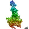
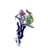
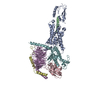
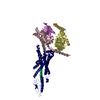
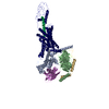
- PDB-5vai: Cryo-EM structure of the activated Glucagon-like peptide-1 recept... -
+ Open data

- Basic information
| Entry | Database: PDB / ID: 5vai | ||||||
|---|---|---|---|---|---|---|---|
| Title | Cryo-EM structure of the activated Glucagon-like peptide-1 receptor in complex with G protein | ||||||
 Components Components |
| ||||||
 Keywords Keywords |  Signaling Protein/Hormone / Class B GPCR / GLP-1 / GLP-1R / Complex / Signaling Protein-Hormone complex Signaling Protein/Hormone / Class B GPCR / GLP-1 / GLP-1R / Complex / Signaling Protein-Hormone complex | ||||||
| Function / homology |  Function and homology informationglucagon receptor binding / glucagon receptor activity / G-protein activation / Activation of the phototransduction cascade / : / Glucagon-type ligand receptors / Thromboxane signalling through TP receptor / Sensory perception of sweet, bitter, and umami (glutamate) taste / G beta:gamma signalling through PI3Kgamma / G beta:gamma signalling through CDC42 ...glucagon receptor binding / glucagon receptor activity / G-protein activation / Activation of the phototransduction cascade / : / Glucagon-type ligand receptors / Thromboxane signalling through TP receptor / Sensory perception of sweet, bitter, and umami (glutamate) taste / G beta:gamma signalling through PI3Kgamma / G beta:gamma signalling through CDC42 / Cooperation of PDCL (PhLP1) and TRiC/CCT in G-protein beta folding / Ca2+ pathway / negative regulation of execution phase of apoptosis / Activation of G protein gated Potassium channels / Synthesis, secretion, and inactivation of Glucagon-like Peptide-1 (GLP-1) / Inhibition of voltage gated Ca2+ channels via Gbeta/gamma subunits / G alpha (z) signalling events / Vasopressin regulates renal water homeostasis via Aquaporins / Glucagon-like Peptide-1 (GLP1) regulates insulin secretion / Adrenaline,noradrenaline inhibits insulin secretion / ADP signalling through P2Y purinoceptor 12 / feeding behavior / G alpha (q) signalling events / G alpha (i) signalling events / Thrombin signalling through proteinase activated receptors (PARs) / Activation of G protein gated Potassium channels / G-protein activation / G beta:gamma signalling through PI3Kgamma / Prostacyclin signalling through prostacyclin receptor / Adrenaline,noradrenaline inhibits insulin secretion / G beta:gamma signalling through PLC beta / ADP signalling through P2Y purinoceptor 1 / Thromboxane signalling through TP receptor / Presynaptic function of Kainate receptors / G beta:gamma signalling through CDC42 / Inhibition of voltage gated Ca2+ channels via Gbeta/gamma subunits / Glucagon-type ligand receptors / alkylglycerophosphoethanolamine phosphodiesterase activity / G alpha (12/13) signalling events / G beta:gamma signalling through BTK / ADP signalling through P2Y purinoceptor 12 / : / Cooperation of PDCL (PhLP1) and TRiC/CCT in G-protein beta folding / Thrombin signalling through proteinase activated receptors (PARs) / Ca2+ pathway / Extra-nuclear estrogen signaling / G alpha (z) signalling events / G alpha (s) signalling events / G alpha (q) signalling events / photoreceptor outer segment membrane / G alpha (i) signalling events / Glucagon-like Peptide-1 (GLP1) regulates insulin secretion / spectrin binding / Vasopressin regulates renal water homeostasis via Aquaporins / positive regulation of calcium ion import / cellular response to glucagon stimulus / positive regulation of insulin secretion involved in cellular response to glucose stimulus / PKA activation in glucagon signalling / regulation of insulin secretion / hair follicle placode formation / intracellular transport / photoreceptor outer segment / D1 dopamine receptor binding / developmental growth / Synthesis, secretion, and deacylation of Ghrelin / Hedgehog 'off' state / positive regulation of cAMP-mediated signaling / adenylate cyclase-activating adrenergic receptor signaling pathway / protein kinase A signaling / positive regulation of gluconeogenesis / cardiac muscle cell apoptotic process / activation of adenylate cyclase activity / photoreceptor inner segment / adenylate cyclase activator activity / response to activity / trans-Golgi network membrane / positive regulation of peptidyl-threonine phosphorylation / gluconeogenesis / G-protein beta/gamma-subunit complex binding / adenylate cyclase-modulating G protein-coupled receptor signaling pathway / bone development / Prostacyclin signalling through prostacyclin receptor / Glucagon signaling in metabolic regulation / adenylate cyclase-activating G protein-coupled receptor signaling pathway / hormone activity / Synthesis, secretion, and inactivation of Glucagon-like Peptide-1 (GLP-1) / platelet aggregation / Glucagon-type ligand receptors / cognition / Vasopressin regulates renal water homeostasis via Aquaporins / G alpha (z) signalling events / positive regulation of GTPase activity / cellular response to catecholamine stimulus / Glucagon-like Peptide-1 (GLP1) regulates insulin secretion / ADORA2B mediated anti-inflammatory cytokines production / adenylate cyclase-activating dopamine receptor signaling pathway / cellular response to prostaglandin E stimulus / sensory perception of taste / GPER1 signaling / G-protein beta-subunit binding Function and homology informationglucagon receptor binding / glucagon receptor activity / G-protein activation / Activation of the phototransduction cascade / : / Glucagon-type ligand receptors / Thromboxane signalling through TP receptor / Sensory perception of sweet, bitter, and umami (glutamate) taste / G beta:gamma signalling through PI3Kgamma / G beta:gamma signalling through CDC42 ...glucagon receptor binding / glucagon receptor activity / G-protein activation / Activation of the phototransduction cascade / : / Glucagon-type ligand receptors / Thromboxane signalling through TP receptor / Sensory perception of sweet, bitter, and umami (glutamate) taste / G beta:gamma signalling through PI3Kgamma / G beta:gamma signalling through CDC42 / Cooperation of PDCL (PhLP1) and TRiC/CCT in G-protein beta folding / Ca2+ pathway / negative regulation of execution phase of apoptosis / Activation of G protein gated Potassium channels / Synthesis, secretion, and inactivation of Glucagon-like Peptide-1 (GLP-1) / Inhibition of voltage gated Ca2+ channels via Gbeta/gamma subunits / G alpha (z) signalling events / Vasopressin regulates renal water homeostasis via Aquaporins / Glucagon-like Peptide-1 (GLP1) regulates insulin secretion / Adrenaline,noradrenaline inhibits insulin secretion / ADP signalling through P2Y purinoceptor 12 / feeding behavior / G alpha (q) signalling events / G alpha (i) signalling events / Thrombin signalling through proteinase activated receptors (PARs) / Activation of G protein gated Potassium channels / G-protein activation / G beta:gamma signalling through PI3Kgamma / Prostacyclin signalling through prostacyclin receptor / Adrenaline,noradrenaline inhibits insulin secretion / G beta:gamma signalling through PLC beta / ADP signalling through P2Y purinoceptor 1 / Thromboxane signalling through TP receptor / Presynaptic function of Kainate receptors / G beta:gamma signalling through CDC42 / Inhibition of voltage gated Ca2+ channels via Gbeta/gamma subunits / Glucagon-type ligand receptors / alkylglycerophosphoethanolamine phosphodiesterase activity / G alpha (12/13) signalling events / G beta:gamma signalling through BTK / ADP signalling through P2Y purinoceptor 12 / : / Cooperation of PDCL (PhLP1) and TRiC/CCT in G-protein beta folding / Thrombin signalling through proteinase activated receptors (PARs) / Ca2+ pathway / Extra-nuclear estrogen signaling / G alpha (z) signalling events / G alpha (s) signalling events / G alpha (q) signalling events / photoreceptor outer segment membrane / G alpha (i) signalling events / Glucagon-like Peptide-1 (GLP1) regulates insulin secretion / spectrin binding / Vasopressin regulates renal water homeostasis via Aquaporins / positive regulation of calcium ion import / cellular response to glucagon stimulus / positive regulation of insulin secretion involved in cellular response to glucose stimulus / PKA activation in glucagon signalling / regulation of insulin secretion / hair follicle placode formation / intracellular transport / photoreceptor outer segment / D1 dopamine receptor binding / developmental growth / Synthesis, secretion, and deacylation of Ghrelin / Hedgehog 'off' state / positive regulation of cAMP-mediated signaling / adenylate cyclase-activating adrenergic receptor signaling pathway / protein kinase A signaling / positive regulation of gluconeogenesis / cardiac muscle cell apoptotic process / activation of adenylate cyclase activity / photoreceptor inner segment / adenylate cyclase activator activity / response to activity / trans-Golgi network membrane / positive regulation of peptidyl-threonine phosphorylation / gluconeogenesis / G-protein beta/gamma-subunit complex binding / adenylate cyclase-modulating G protein-coupled receptor signaling pathway / bone development / Prostacyclin signalling through prostacyclin receptor / Glucagon signaling in metabolic regulation / adenylate cyclase-activating G protein-coupled receptor signaling pathway / hormone activity / Synthesis, secretion, and inactivation of Glucagon-like Peptide-1 (GLP-1) / platelet aggregation / Glucagon-type ligand receptors / cognition / Vasopressin regulates renal water homeostasis via Aquaporins / G alpha (z) signalling events / positive regulation of GTPase activity / cellular response to catecholamine stimulus / Glucagon-like Peptide-1 (GLP1) regulates insulin secretion / ADORA2B mediated anti-inflammatory cytokines production / adenylate cyclase-activating dopamine receptor signaling pathway / cellular response to prostaglandin E stimulus / sensory perception of taste / GPER1 signaling / G-protein beta-subunit bindingSimilarity search - Function | ||||||
| Biological species |  Oryctolagus cuniculus (rabbit) Oryctolagus cuniculus (rabbit) Homo sapiens (human)Rattus norvegicus (Norway rat)Bos taurus (cattle)Lama glama (llama) Homo sapiens (human)Rattus norvegicus (Norway rat)Bos taurus (cattle)Lama glama (llama) | ||||||
| Method | ELECTRON MICROSCOPY / single particle reconstruction / cryo EM / Resolution: 4.1 Å | ||||||
 Authors Authors | Zhang, Y. / Sun, B. / Feng, D. / Hu, H. / Chu, M. / Qu, Q. / Tarrasch, J.T. / Li, S. / Kobilka, T.S. / Kobilka, B.K. / Skiniotis, G. | ||||||
 Citation Citation | Journal: Nature / Year: 2017 Title: Cryo-EM structure of the activated GLP-1 receptor in complex with a G protein. Authors: Yan Zhang / Bingfa Sun / Dan Feng / Hongli Hu / Matthew Chu / Qianhui Qu / Jeffrey T Tarrasch / Shane Li / Tong Sun Kobilka / Brian K Kobilka / Georgios Skiniotis /  Abstract: Glucagon-like peptide 1 (GLP-1) is a hormone with essential roles in regulating insulin secretion, carbohydrate metabolism and appetite. GLP-1 effects are mediated through binding to the GLP-1 ...Glucagon-like peptide 1 (GLP-1) is a hormone with essential roles in regulating insulin secretion, carbohydrate metabolism and appetite. GLP-1 effects are mediated through binding to the GLP-1 receptor (GLP-1R), a class B G-protein-coupled receptor (GPCR) that signals primarily through the stimulatory G protein G. Class B GPCRs are important therapeutic targets; however, our understanding of their mechanism of action is limited by the lack of structural information on activated and full-length receptors. Here we report the cryo-electron microscopy structure of the peptide-activated GLP-1R-G complex at near atomic resolution. The peptide is clasped between the N-terminal domain and the transmembrane core of the receptor, and further stabilized by extracellular loops. Conformational changes in the transmembrane domain result in a sharp kink in the middle of transmembrane helix 6, which pivots its intracellular half outward to accommodate the α5-helix of the Ras-like domain of G. These results provide a structural framework for understanding class B GPCR activation through hormone binding. | ||||||
| History |
|
- Structure visualization
| Movie |
Movie viewer |
|---|---|
| Structure viewer | Molecule: MolmilJmol/JSmol |
- Downloads & links
-Download
| PDBx/mmCIF format | 5vai.cif.gz | 218.9 KB | Display | PDBx/mmCIF format |
|---|---|---|---|---|
| PDB format | pdb5vai.ent.gz | 172.2 KB | Display | PDB format |
| PDBx/mmJSON format | 5vai.json.gz | Tree view | PDBx/mmJSON format | |
| Others |  Other downloads Other downloads |
-Validation report
| Arichive directory | https://data.pdbj.org/pub/pdb/validation_reports/va/5vaiftp://data.pdbj.org/pub/pdb/validation_reports/va/5vai | HTTPS FTP |
|---|
-Related structure data
| Related structure data | 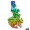 8653MC M: map data used to model this data C: citing same article ( |
|---|---|
| Similar structure data |


-Links
 PDBj
PDBj


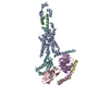
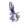
- Assembly
| Deposited unit | 
|
|---|---|
| 1 |
|
-Components
-Guanine nucleotide-binding protein ... , 3 types, 3 molecules ABG
| #3: Protein | Mass: 44370.168 Da / Num. of mol.: 1 / Fragment: UNP residuews 1-380 Source method: isolated from a genetically manipulated source Source: (gene. exp.) Homo sapiens (human) / Gene: GNAS, GNAS1, GSP / Production host:  Trichoplusia ni (cabbage looper) / References: UniProt: P63092 Trichoplusia ni (cabbage looper) / References: UniProt: P63092 |
|---|---|
| #4: Protein | Mass: 38744.371 Da / Num. of mol.: 1 Source method: isolated from a genetically manipulated source Source: (gene. exp.) Rattus norvegicus (Norway rat) / Gene: Gnb1 / Production host: Trichoplusia ni (cabbage looper) / References: UniProt: P54311 |
| #5: Protein | Mass: 7563.750 Da / Num. of mol.: 1 / Fragment: UNP residues 1-68 Source method: isolated from a genetically manipulated source Source: (gene. exp.) Bos taurus (cattle) / Gene: GNG2 / Production host: Trichoplusia ni (cabbage looper) / References: UniProt: P63212 |
-Protein / Protein/peptide / Antibody , 3 types, 3 molecules RPN
| #1: Protein | Mass: 52581.793 Da / Num. of mol.: 1 Source method: isolated from a genetically manipulated source Source: (gene. exp.) Oryctolagus cuniculus (rabbit) / Gene: GLP1R / Production host:  Spodoptera frugiperda (fall armyworm) / References: UniProt: G1SGD4 Spodoptera frugiperda (fall armyworm) / References: UniProt: G1SGD4 |
|---|---|
| #2: Protein/peptide | Glucagon-like peptide-1 Mass: 3359.699 Da / Num. of mol.: 1 / Fragment: UNP residues 98-128 / Source method: obtained synthetically / Source: (synth.) Homo sapiens (human) / References: UniProt: P01275 |
| #6: Antibody | Single-domain antibody Mass: 15140.742 Da / Num. of mol.: 1 Source method: isolated from a genetically manipulated source Source: (gene. exp.) Lama glama (llama) / Production host:  Escherichia coli (E. coli) Escherichia coli (E. coli) |
-Experimental details
-Experiment
| Experiment | Method: ELECTRON MICROSCOPY |
|---|---|
| EM experiment | Aggregation state: PARTICLE / 3D reconstruction method: single particle reconstruction |
- Sample preparation
| Component | Name: the activated glucagon-like peptide-1 receptor in complex with the heterotrimeric Gs protein Type: COMPLEX / Entity ID: all / Source: RECOMBINANT |
|---|---|
| Molecular weight | Value: 0.15 MDa / Experimental value: NO |
| Source (natural) | Organism: Oryctolagus cuniculus (rabbit) |
| Source (recombinant) | Organism: Spodoptera frugiperda (fall armyworm) |
| Buffer solution | pH: 7.5 |
| Specimen | Conc.: 1 mg/ml / Embedding applied: NO / Shadowing applied: NO / Staining applied: NO / Vitrification applied: YES |
| Specimen support | Grid type: Quantifoil |
| Vitrification | Instrument: FEI VITROBOT MARK IV / Cryogen name: ETHANE / Humidity: 100 % |
- Electron microscopy imaging
| Experimental equipment |  Model: Titan Krios / Image courtesy: FEI Company |
|---|---|
| Microscopy | Model: FEI TITAN KRIOS |
| Electron gun | Electron source: FIELD EMISSION GUN / Accelerating voltage: 300 kV / Illumination mode: FLOOD BEAM |
| Electron lens | Mode: BRIGHT FIELDBright-field microscopy / Nominal magnification: 29000 X / Calibrated magnification: 50000 X / Alignment procedure: COMA FREE |
| Specimen holder | Cryogen: NITROGEN / Specimen holder model: FEI TITAN KRIOS AUTOGRID HOLDER |
| Image recording | Electron dose: 90 e/Å2 / Detector mode: COUNTING / Film or detector model: GATAN K2 SUMMIT (4k x 4k) |
- Processing
| Software | Name: PHENIX / Version: 1.11.1_2575: / Classification: refinement | ||||||||||||||||||||||||
|---|---|---|---|---|---|---|---|---|---|---|---|---|---|---|---|---|---|---|---|---|---|---|---|---|---|
| EM software |
| ||||||||||||||||||||||||
| CTF correction | Type: PHASE FLIPPING AND AMPLITUDE CORRECTION | ||||||||||||||||||||||||
| Symmetry | Point symmetry: C1 (asymmetric) | ||||||||||||||||||||||||
| 3D reconstruction | Resolution: 4.1 Å / Resolution method: FSC 0.143 CUT-OFF / Num. of particles: 139299 / Algorithm: BACK PROJECTION / Num. of class averages: 1 / Symmetry type: POINT | ||||||||||||||||||||||||
| Refine LS restraints |
|
