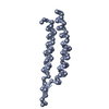
Journal: Nat Commun / Year: 2017 Title: Unique architecture of thermophilic archaeal virus APBV1 and its genome packaging. Authors: Denis Ptchelkine / Ashley Gillum / Tomohiro Mochizuki / Soizick Lucas-Staat / Ying Liu / Mart Krupovic / Simon E V Phillips / David Prangishvili / Juha T Huiskonen / Abstract: Archaeal viruses have evolved to infect hosts often thriving in extreme conditions such as high temperatures. However, there is a paucity of information on archaeal virion structures, genome ...Archaeal viruses have evolved to infect hosts often thriving in extreme conditions such as high temperatures. However, there is a paucity of information on archaeal virion structures, genome packaging, and determinants of temperature resistance. The rod-shaped virus APBV1 (Aeropyrum pernix bacilliform virus 1) is among the most thermostable viruses known; it infects a hyperthermophile Aeropyrum pernix, which grows optimally at 90 °C. Here we report the structure of APBV1, determined by cryo-electron microscopy at near-atomic resolution. Tight packing of the major virion glycoprotein (VP1) is ensured by extended hydrophobic interfaces, and likely contributes to the extreme thermostability of the helical capsid. The double-stranded DNA is tightly packed in the capsid as a left-handed superhelix and held in place by the interactions with positively charged residues of VP1. The assembly is closed by specific capping structures at either end, which we propose to play a role in DNA packing and delivery.
History
Deposition
Sep 6, 2017
Deposition site: PDBE / Processing site: PDBE
Revision 1.0
Nov 22, 2017
Provider: repository / Type: Initial release
Revision 1.1
Oct 17, 2018
Group: Data collection / Refinement description / Category: refine
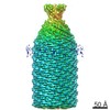
Num. of particles selected: 169316 Details: Overlapping segments extracted from helical particles
Symmetry
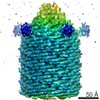
Point symmetry: C5 (5 fold cyclic)
3D reconstruction
Resolution: 3.7 Å / Resolution method: FSC 0.143 CUT-OFF / Num. of particles: 94645 / Algorithm: FOURIER SPACE / Symmetry type: POINT
Atomic model building
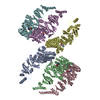
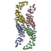
Protocol: AB INITIO MODEL
Refinement
Resolution: 3.5→113.4 Å / Cor.coef. Fo:Fc: 0.898 / ESU R: 0.328 Stereochemistry target values: MAXIMUM LIKELIHOOD WITH PHASES Details: HYDROGENS HAVE BEEN ADDED IN THE RIDING POSITIONS
Rfactor
Num. reflection
% reflection
Rwork
0.27071
-
-
obs
0.27071
43610
100 %
Solvent computation
Ion probe radii: 0.8 Å / Shrinkage radii: 0.8 Å / VDW probe radii: 1.2 Å / Solvent model: MASK
 Movie
Movie Controller

 Yorodumi
 Open data
 Basic information
 Components
 Keywords
 VIRUS /
  Function and homology information

 Authors
 United Kingdom, 4items
  Citation


 Structure visualization
 Downloads & links
 Other downloads


 PDBj
PDBj Assembly
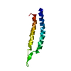
 Sample preparation
 Electron microscopy imaging
 Processing