 Movie
Movie Controller

[English] 日本語
 Yorodumi
- PDB-5np7: CryoEM structure of Human Rad51 on single-stranded DNA to 4.2A re... -
+ Open data

- Basic information
| Entry | Database: PDB / ID: 5np7 | ||||||
|---|---|---|---|---|---|---|---|
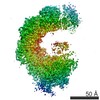
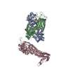
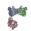
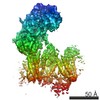
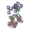
| Title | CryoEM structure of Human Rad51 on single-stranded DNA to 4.2A resolution. | ||||||
 Components Components | DNA repair protein RAD51 homolog 1 | ||||||
 Keywords Keywords | RECOMBINATION / recombinase / cryoEM / Human Rad51 / single-stranded DNA | ||||||
| Function / homology |  Function and homology information Function and homology informationpresynaptic intermediate filament cytoskeleton / mitotic recombination-dependent replication fork processing / chromosome organization involved in meiotic cell cycle / cellular response to camptothecin / DNA recombinase assembly / telomere maintenance via telomere lengthening / positive regulation of DNA ligation / mitotic recombination / double-strand break repair involved in meiotic recombination / nuclear ubiquitin ligase complex ...presynaptic intermediate filament cytoskeleton / mitotic recombination-dependent replication fork processing / chromosome organization involved in meiotic cell cycle / cellular response to camptothecin / DNA recombinase assembly / telomere maintenance via telomere lengthening / positive regulation of DNA ligation / mitotic recombination / double-strand break repair involved in meiotic recombination / nuclear ubiquitin ligase complex / DNA strand invasion / replication-born double-strand break repair via sister chromatid exchange / cellular response to hydroxyurea / DNA strand exchange activity / lateral element / telomere maintenance via recombination / regulation of DNA damage checkpoint / Impaired BRCA2 binding to PALB2 / single-stranded DNA helicase activity / reciprocal meiotic recombination / Defective homologous recombination repair (HRR) due to BRCA1 loss of function / Defective HDR through Homologous Recombination Repair (HRR) due to PALB2 loss of BRCA1 binding function / Defective HDR through Homologous Recombination Repair (HRR) due to PALB2 loss of BRCA2/RAD51/RAD51C binding function / Homologous DNA Pairing and Strand Exchange / Resolution of D-loop Structures through Synthesis-Dependent Strand Annealing (SDSA) / Resolution of D-loop Structures through Holliday Junction Intermediates / HDR through Single Strand Annealing (SSA) / Impaired BRCA2 binding to RAD51 / ATP-dependent DNA damage sensor activity / regulation of double-strand break repair via homologous recombination / nuclear chromosome / replication fork processing / DNA unwinding involved in DNA replication / Transcriptional Regulation by E2F6 / Presynaptic phase of homologous DNA pairing and strand exchange / ATP-dependent activity, acting on DNA / interstrand cross-link repair / DNA polymerase binding / condensed chromosome / meiotic cell cycle / condensed nuclear chromosome / male germ cell nucleus / cellular response to ionizing radiation / double-strand break repair via homologous recombination / regulation of protein phosphorylation / HDR through Homologous Recombination (HRR) / PML body / Meiotic recombination / single-stranded DNA binding / site of double-strand break / double-stranded DNA binding / DNA recombination / chromosome, telomeric region / mitochondrial matrix / DNA repair / centrosome / DNA damage response / chromatin binding / chromatin / nucleolus / perinuclear region of cytoplasm / enzyme binding / ATP hydrolysis activity / protein-containing complex / mitochondrion / nucleoplasm / ATP binding / identical protein binding / nucleus / cytosol / cytoplasmSimilarity search - Function | ||||||
| Biological species |  Homo sapiens (human) Homo sapiens (human) | ||||||
| Method | ELECTRON MICROSCOPY / helical reconstruction / cryo EM / Resolution: 4.2 Å | ||||||
 Authors Authors | Short, J.M. / Venkitaraman, A. | ||||||
 Citation Citation | Journal: Nucleic Acids Res / Year: 2016 Title: High-resolution structure of the presynaptic RAD51 filament on single-stranded DNA by electron cryo-microscopy. Authors: Judith M Short / Yang Liu / Shaoxia Chen / Neelesh Soni / Mallur S Madhusudhan / Mahmud K K Shivji / Ashok R Venkitaraman /    Abstract: Homologous DNA recombination (HR) by the RAD51 recombinase enables error-free DNA break repair. To execute HR, RAD51 first forms a presynaptic filament on single-stranded (ss) DNA, which catalyses ...Homologous DNA recombination (HR) by the RAD51 recombinase enables error-free DNA break repair. To execute HR, RAD51 first forms a presynaptic filament on single-stranded (ss) DNA, which catalyses pairing with homologous double-stranded (ds) DNA. Here, we report a structure for the presynaptic human RAD51 filament at 3.5-5.0Å resolution using electron cryo-microscopy. RAD51 encases ssDNA in a helical filament of 103Å pitch, comprising 6.4 protomers per turn, with a rise of 16.1Å and a twist of 56.2°. Inter-protomer distance correlates with rotation of an α-helical region in the core catalytic domain that is juxtaposed to ssDNA, suggesting how the RAD51-DNA interaction modulates protomer spacing and filament pitch. We map Fanconi anaemia-like disease-associated RAD51 mutations, clarifying potential phenotypes. We predict binding sites on the presynaptic filament for two modules present in each BRC repeat of the BRCA2 tumour suppressor, a critical HR mediator. Structural modelling suggests that changes in filament pitch mask or expose one binding site with filament-inhibitory potential, rationalizing the paradoxical ability of the BRC repeats to either stabilize or inhibit filament formation at different steps during HR. Collectively, our findings provide fresh insight into the structural mechanism of HR and its dysregulation in human disease. | ||||||
| History |
|
- Structure visualization
| Movie |
Movie viewer |
|---|---|
| Structure viewer | Molecule: MolmilJmol/JSmol |
- Downloads & links
-Download
| PDBx/mmCIF format | 5np7.cif.gz | 416.3 KB | Display | PDBx/mmCIF format |
|---|---|---|---|---|
| PDB format | pdb5np7.ent.gz | 346.4 KB | Display | PDB format |
| PDBx/mmJSON format | 5np7.json.gz | Tree view | PDBx/mmJSON format | |
| Others |  Other downloads Other downloads |
-Validation report
| Arichive directory | https://data.pdbj.org/pub/pdb/validation_reports/np/5np7ftp://data.pdbj.org/pub/pdb/validation_reports/np/5np7 | HTTPS FTP |
|---|
-Related structure data
| Related structure data | 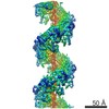 8183MC 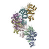 5jzcC M: map data used to model this data C: citing same article ( |
|---|---|
| Similar structure data |


-Links
 PDBj
PDBj


- Assembly
| Deposited unit | 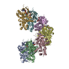
|
|---|---|
| 1 |
|
-Components
| #1: Protein | / hRAD51 / RAD51 homolog A Mass: 37009.125 Da / Num. of mol.: 7 Source method: isolated from a genetically manipulated source Source: (gene. exp.) Homo sapiens (human) / Gene: RAD51, RAD51A, RECA / Production host:  Escherichia coli (E. coli) / References: UniProt: Q06609 Escherichia coli (E. coli) / References: UniProt: Q06609#2: Chemical | ChemComp-ANP /   Mass: 506.196 Da / Num. of mol.: 7 / Source method: obtained synthetically / Formula: C10H17N6O12P3 / Comment: AMP-PNP, energy-carrying molecule analogue*YM Mass: 506.196 Da / Num. of mol.: 7 / Source method: obtained synthetically / Formula: C10H17N6O12P3 / Comment: AMP-PNP, energy-carrying molecule analogue*YM |
|---|
-Experimental details
-Experiment
| Experiment | Method: ELECTRON MICROSCOPY |
|---|---|
| EM experiment | Aggregation state: HELICAL ARRAY / 3D reconstruction method: helical reconstruction |
- Sample preparation
| Component | Name: Helical filament of HRAD51 on single-stranded DNA with AMPPNP Type: COMPLEX / Entity ID: #1 / Source: RECOMBINANT |
|---|---|
| Molecular weight | Value: 259 kDa/nm / Experimental value: NO |
| Source (natural) | Organism: Homo sapiens (human) |
| Source (recombinant) | Organism: Escherichia coli (E. coli) |
| Buffer solution | pH: 7 |
| Specimen | Embedding applied: NO / Shadowing applied: NO / Staining applied: NO / Vitrification applied: YES |
| Specimen support | Grid material: COPPER / Grid mesh size: 400 divisions/in. / Grid type: Quantifoil |
| Vitrification | Cryogen name: NITROGEN |
- Electron microscopy imaging
| Experimental equipment |  Model: Titan Krios / Image courtesy: FEI Company |
|---|---|
| Microscopy | Model: FEI TITAN KRIOS |
| Electron gun | Electron source: FIELD EMISSION GUN / Accelerating voltage: 300 kV / Illumination mode: FLOOD BEAM |
| Electron lens | Mode: BRIGHT FIELDBright-field microscopy / Nominal magnification: 59000 X / Calibrated magnification: 104477 X / Nominal defocus min: 2000 nm / Cs: 2.7 mm / Alignment procedure: COMA FREE |
| Specimen holder | Cryogen: NITROGEN / Specimen holder model: FEI TITAN KRIOS AUTOGRID HOLDER |
| Image recording | Electron dose: 20 e/Å2 / Detector mode: INTEGRATING / Film or detector model: FEI FALCON II (4k x 4k) |
| Image scans | Sampling size: 14 µm / Width: 4096 / Height: 4096 / Movie frames/image: 34 |
- Processing
| Software | Name: REFMAC / Version: 5.8.0166 / Classification: refinement | ||||||||||||||||||||||||||||||||||||||||||||||||||||||||||||||||||||||||||||||||||||||||||||||||||||||||||
|---|---|---|---|---|---|---|---|---|---|---|---|---|---|---|---|---|---|---|---|---|---|---|---|---|---|---|---|---|---|---|---|---|---|---|---|---|---|---|---|---|---|---|---|---|---|---|---|---|---|---|---|---|---|---|---|---|---|---|---|---|---|---|---|---|---|---|---|---|---|---|---|---|---|---|---|---|---|---|---|---|---|---|---|---|---|---|---|---|---|---|---|---|---|---|---|---|---|---|---|---|---|---|---|---|---|---|---|
| EM software |
| ||||||||||||||||||||||||||||||||||||||||||||||||||||||||||||||||||||||||||||||||||||||||||||||||||||||||||
| CTF correction | Details: per segment / Type: NONE | ||||||||||||||||||||||||||||||||||||||||||||||||||||||||||||||||||||||||||||||||||||||||||||||||||||||||||
| Helical symmerty | Angular rotation/subunit: 56.2 ° / Axial rise/subunit: 16 Å / Axial symmetry: C1 | ||||||||||||||||||||||||||||||||||||||||||||||||||||||||||||||||||||||||||||||||||||||||||||||||||||||||||
| Particle selection | Num. of particles selected: 70000 | ||||||||||||||||||||||||||||||||||||||||||||||||||||||||||||||||||||||||||||||||||||||||||||||||||||||||||
| 3D reconstruction | Resolution: 4.2 Å / Resolution method: FSC 0.143 CUT-OFF / Num. of particles: 60000 Details: Helical segments were divided in half, not drawn randomly or odd/even as this would result in some segments from the same filament in both half models. Thus, it is strictly not gold-standard. Symmetry type: HELICAL | ||||||||||||||||||||||||||||||||||||||||||||||||||||||||||||||||||||||||||||||||||||||||||||||||||||||||||
| Atomic model building | B value: 94.574 / Protocol: FLEXIBLE FIT / Space: RECIPROCAL / Target criteria: Maximum likelihood Details: Backbone fitting is good but many of the sidechains cannot be relied on as the map resolution was insufficient. | ||||||||||||||||||||||||||||||||||||||||||||||||||||||||||||||||||||||||||||||||||||||||||||||||||||||||||
| Refinement | Resolution: 4.2→180 Å / Cor.coef. Fo:Fc: 0.937 / SU B: 46.567 / SU ML: 0.572 Stereochemistry target values: MAXIMUM LIKELIHOOD WITH PHASES
| ||||||||||||||||||||||||||||||||||||||||||||||||||||||||||||||||||||||||||||||||||||||||||||||||||||||||||
| Solvent computation | Solvent model: PARAMETERS FOR MASK CACLULATION | ||||||||||||||||||||||||||||||||||||||||||||||||||||||||||||||||||||||||||||||||||||||||||||||||||||||||||
| Displacement parameters | Biso mean: 131.509 Å2
| ||||||||||||||||||||||||||||||||||||||||||||||||||||||||||||||||||||||||||||||||||||||||||||||||||||||||||
| Refinement step | Cycle: 1 / Total: 16754 | ||||||||||||||||||||||||||||||||||||||||||||||||||||||||||||||||||||||||||||||||||||||||||||||||||||||||||
| Refine LS restraints |
|