 Movie
Movie Controller

[English] 日本語
 Yorodumi
- PDB-5mz6: Cryo-EM structure of a Separase-Securin complex from Caenorhabdit... -
+ Open data

- Basic information
| Entry | Database: PDB / ID: 5mz6 | ||||||
|---|---|---|---|---|---|---|---|
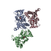
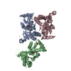
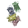
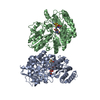
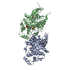
| Title | Cryo-EM structure of a Separase-Securin complex from Caenorhabditis elegans at 3.8 A resolution | ||||||
 Components Components |
| ||||||
 Keywords Keywords |  CELL CYCLE / caspase / cohesin / cleavage CELL CYCLE / caspase / cohesin / cleavage | ||||||
| Function / homology |  Function and homology information Function and homology informationSeparation of Sister Chromatids / separase-securin complex / eggshell formation / metaphase plate / regulation of nematode larval development / separase / cortical granule exocytosis / maintenance of meiotic sister chromatid cohesion / meiotic chromosome separation / maintenance of mitotic sister chromatid cohesion ...Separation of Sister Chromatids / separase-securin complex / eggshell formation / metaphase plate / regulation of nematode larval development / separase / cortical granule exocytosis / maintenance of meiotic sister chromatid cohesion / meiotic chromosome separation / maintenance of mitotic sister chromatid cohesion / polarity specification of anterior/posterior axis / polar body extrusion after meiotic divisions / cortical granule / regulation of centriole-centriole cohesion / mitotic sister chromatid separation / regulation of exocytosis / multicellular organismal reproductive process / meiotic spindle / regulation of locomotion / centrosome localization / cleavage furrow / centrosome duplication / mitotic cytokinesis / condensed chromosome / condensed nuclear chromosome / spindle microtubule / protein localization / spindle / mitotic spindle / protein processing / nuclear envelope / chromosome / cell cortex / midbody / protease binding / protein stabilization / cysteine-type endopeptidase activity / centrosome / ubiquitin protein ligase binding / nucleus / cytoplasmSimilarity search - Function | ||||||
| Biological species |  Caenorhabditis elegans (invertebrata) Caenorhabditis elegans (invertebrata) | ||||||
| Method | ELECTRON MICROSCOPY / single particle reconstruction / cryo EM / Resolution: 3.8 Å | ||||||
 Authors Authors | Boland, A. / Martin, T.G. / Zhang, Z. / Yang, J. / Bai, X.C. / Chang, L. / Scheres, S.H.W. / Barford, D. | ||||||
 Citation Citation | Journal: Nat Struct Mol Biol / Year: 2017 Title: Cryo-EM structure of a metazoan separase-securin complex at near-atomic resolution. Authors: Andreas Boland / Thomas G Martin / Ziguo Zhang / Jing Yang / Xiao-Chen Bai / Leifu Chang / Sjors H W Scheres / David Barford /  Abstract: Separase is a caspase-family protease that initiates chromatid segregation by cleaving the kleisin subunits (Scc1 and Rec8) of cohesin, and regulates centrosome duplication and mitotic spindle ...Separase is a caspase-family protease that initiates chromatid segregation by cleaving the kleisin subunits (Scc1 and Rec8) of cohesin, and regulates centrosome duplication and mitotic spindle function through cleavage of kendrin and Slk19. To understand the mechanisms of securin regulation of separase, we used single-particle cryo-electron microscopy (cryo-EM) to determine a near-atomic-resolution structure of the Caenorhabditis elegans separase-securin complex. Separase adopts a triangular-shaped bilobal architecture comprising an N-terminal tetratricopeptide repeat (TPR)-like α-solenoid domain docked onto the conserved C-terminal protease domain. Securin engages separase in an extended antiparallel conformation, interacting with both lobes. It inhibits separase by interacting with the catalytic site through a pseudosubstrate mechanism, thus revealing that in the inhibited separase-securin complex, the catalytic site adopts a conformation compatible with substrate binding. Securin is protected from cleavage because an aliphatic side chain at the P1 position represses protease activity by disrupting the organization of catalytic site residues. | ||||||
| History |
|
- Structure visualization
| Movie |
Movie viewer |
|---|---|
| Structure viewer | Molecule: MolmilJmol/JSmol |
- Downloads & links
-Download
| PDBx/mmCIF format | 5mz6.cif.gz | 202.7 KB | Display | PDBx/mmCIF format |
|---|---|---|---|---|
| PDB format | pdb5mz6.ent.gz | 160.1 KB | Display | PDB format |
| PDBx/mmJSON format | 5mz6.json.gz | Tree view | PDBx/mmJSON format | |
| Others |  Other downloads Other downloads |
-Validation report
| Arichive directory | https://data.pdbj.org/pub/pdb/validation_reports/mz/5mz6ftp://data.pdbj.org/pub/pdb/validation_reports/mz/5mz6 | HTTPS FTP |
|---|
-Related structure data
| Related structure data | 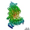 3583MC  3584C M: map data used to model this data C: citing same article ( |
|---|---|
| Similar structure data |


-Links
 PDBj
PDBj
- Assembly
| Deposited unit | 
|
|---|---|
| 1 |
|
-Components
| #1: Protein | / Separase Mass: 144315.984 Da / Num. of mol.: 1 Source method: isolated from a genetically manipulated source Source: (gene. exp.) Caenorhabditis elegans (invertebrata) / Gene: sep-1, CELE_Y47G6A.12, Y47G6A.12 / Production host:  Trichoplusia ni (cabbage looper) / References: UniProt: G5ED39 Trichoplusia ni (cabbage looper) / References: UniProt: G5ED39 |
|---|---|
| #2: Protein | Mass: 27027.646 Da / Num. of mol.: 1 Source method: isolated from a genetically manipulated source Source: (gene. exp.) Caenorhabditis elegans (invertebrata) / Gene: ify-1, C27A2.3, CELE_C27A2.3 / Production host: Trichoplusia ni (cabbage looper) / References: UniProt: Q18235 |
-Experimental details
-Experiment
| Experiment | Method: ELECTRON MICROSCOPY |
|---|---|
| EM experiment | Aggregation state: PARTICLE / 3D reconstruction method: single particle reconstruction |
- Sample preparation
| Component | Name: Caenorhabditis elegans Separase-Securin complex / Type: COMPLEX Details: C. elegans Separase-Securin complex at 3.8 Angstrom resolution refined with Relion. Entity ID: all / Source: RECOMBINANT |
|---|---|
| Molecular weight | Value: 0.17 MDa / Experimental value: NO |
| Source (natural) | Organism: Caenorhabditis elegans (invertebrata) |
| Source (recombinant) | Organism: Trichoplusia ni (cabbage looper) |
| Buffer solution | pH: 7.8 |
| Specimen | Conc.: 0.02 mg/ml / Embedding applied: NO / Shadowing applied: NO / Staining applied: NO / Vitrification applied: YES / Details: Monodisperse sample |
| Specimen support | Details: Glow discharging was performed before applying graphene oxide solution onto the grid. Grid was washed three times, dried and sample was applied. For detailed information see: https: ...Details: Glow discharging was performed before applying graphene oxide solution onto the grid. Grid was washed three times, dried and sample was applied. For detailed information see: https://figshare.com/articles/Graphene_Oxide_Grid_Preparation/3178669 Grid material: GOLD / Grid mesh size: 300 divisions/in. / Grid type: Quantifoil R1.2/1.3 |
| Vitrification | Cryogen name: ETHANE / Humidity: 100 % / Chamber temperature: 277 K / Details: Custom Manual Plunger |
- Electron microscopy imaging
| Experimental equipment |  Model: Titan Krios / Image courtesy: FEI Company |
|---|---|
| Microscopy | Model: FEI TITAN KRIOS |
| Electron gun | Electron source: FIELD EMISSION GUN / Accelerating voltage: 300 kV / Illumination mode: FLOOD BEAM |
| Electron lens | Mode: BRIGHT FIELDBright-field microscopy / Cs: 2.7 mm / C2 aperture diameter: 70 µm / Alignment procedure: COMA FREE |
| Specimen holder | Cryogen: NITROGEN / Specimen holder model: FEI TITAN KRIOS AUTOGRID HOLDER |
| Image recording | Average exposure time: 0.8 sec. / Electron dose: 1.25 e/Å2 / Detector mode: SUPER-RESOLUTION / Film or detector model: GATAN K2 SUMMIT (4k x 4k) |
| EM imaging optics | Energyfilter name: GIF / Energyfilter upper: 20 eV / Energyfilter lower: -20 eV |
| Image scans | Width: 3710 / Height: 3710 |
- Processing
| Software | Name: PHENIX / Version: 1.11.1_2575: / Classification: refinement | ||||||||||||||||||||||||
|---|---|---|---|---|---|---|---|---|---|---|---|---|---|---|---|---|---|---|---|---|---|---|---|---|---|
| EM software |
| ||||||||||||||||||||||||
| EM 3D crystal entity | ∠α: 90 ° / ∠β: 90 ° / ∠γ: 90 ° / A: 1 Å / B: 1 Å / C: 1 Å / Space group name: 1 / Space group num: 1 | ||||||||||||||||||||||||
| CTF correction | Type: PHASE FLIPPING AND AMPLITUDE CORRECTION | ||||||||||||||||||||||||
| Symmetry | Point symmetry: C1 (asymmetric) | ||||||||||||||||||||||||
| 3D reconstruction | Resolution: 3.8 Å / Resolution method: FSC 0.143 CUT-OFF / Num. of particles: 103696 / Symmetry type: POINT | ||||||||||||||||||||||||
| Atomic model building | Protocol: AB INITIO MODEL / Space: REAL | ||||||||||||||||||||||||
| Refine LS restraints |
|
