 Movie
Movie Controller

[English] 日本語
 Yorodumi
- PDB-5muv: Atomic structure fitted into a localized reconstruction of bacter... -
+ Open data

- Basic information
| Entry | Database: PDB / ID: 5muv | ||||||||||||
|---|---|---|---|---|---|---|---|---|---|---|---|---|---|
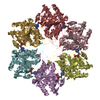
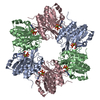
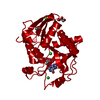
| Title | Atomic structure fitted into a localized reconstruction of bacteriophage phi6 packaging hexamer P4 | ||||||||||||
 Components Components | Packaging enzyme P4 | ||||||||||||
 Keywords Keywords |  HYDROLASE / packaging / ATPase / vertex / hyrdolase HYDROLASE / packaging / ATPase / vertex / hyrdolase | ||||||||||||
| Function / homology |  Function and homology information Function and homology informationviral procapsid / viral genome packaging / ribonucleoside triphosphate phosphatase activity / viral capsid / nucleoside-triphosphate phosphatase / ATP bindingSimilarity search - Function | ||||||||||||
| Biological species |  Pseudomonas phage phi6 (bacteriophage) Pseudomonas phage phi6 (bacteriophage) | ||||||||||||
| Method | ELECTRON MICROSCOPY / single particle reconstruction / cryo EM / Resolution: 9.1 Å | ||||||||||||
 Authors Authors | Sun, Z. / El Omari, K. / Sun, X. / Ilca, S. / Kotecha, A. / Stuart, D.I. / Poranen, M.M. / Huiskonen, J.T. | ||||||||||||
| Funding support |  Finland, 1items Finland, 1items
| ||||||||||||
 Citation Citation | Journal: Nat Commun / Year: 2017 Title: Double-stranded RNA virus outer shell assembly by bona fide domain-swapping. Authors: Zhaoyang Sun / Kamel El Omari / Xiaoyu Sun / Serban L Ilca / Abhay Kotecha / David I Stuart / Minna M Poranen / Juha T Huiskonen /  Abstract: Correct outer protein shell assembly is a prerequisite for virion infectivity in many multi-shelled dsRNA viruses. In the prototypic dsRNA bacteriophage φ6, the assembly reaction is promoted by ...Correct outer protein shell assembly is a prerequisite for virion infectivity in many multi-shelled dsRNA viruses. In the prototypic dsRNA bacteriophage φ6, the assembly reaction is promoted by calcium ions but its biomechanics remain poorly understood. Here, we describe the near-atomic resolution structure of the φ6 double-shelled particle. The outer T=13 shell protein P8 consists of two alpha-helical domains joined by a linker, which allows the trimer to adopt either a closed or an open conformation. The trimers in an open conformation swap domains with each other. Our observations allow us to propose a mechanistic model for calcium concentration regulated outer shell assembly. Furthermore, the structure provides a prime exemplar of bona fide domain-swapping. This leads us to extend the theory of domain-swapping from the level of monomeric subunits and multimers to closed spherical shells, and to hypothesize a mechanism by which closed protein shells may arise in evolution. #1: Journal: Nucleic Acids Res / Year: 2013Title: Tracking in atomic detail the functional specializations in viral RecA helicases that occur during evolution. Authors: Kamel El Omari / Christoph Meier / Denis Kainov / Geoff Sutton / Jonathan M Grimes / Minna M Poranen / Dennis H Bamford / Roman Tuma / David I Stuart / Erika J Mancini / Abstract: Many complex viruses package their genomes into empty protein shells and bacteriophages of the Cystoviridae family provide some of the simplest models for this. The cystoviral hexameric NTPase, P4, ...Many complex viruses package their genomes into empty protein shells and bacteriophages of the Cystoviridae family provide some of the simplest models for this. The cystoviral hexameric NTPase, P4, uses chemical energy to translocate single-stranded RNA genomic precursors into the procapsid. We previously dissected the mechanism of RNA translocation for one such phage, 12, and have now investigated three further highly divergent, cystoviral P4 NTPases (from 6, 8 and 13). High-resolution crystal structures of the set of P4s allow a structure-based phylogenetic analysis, which reveals that these proteins form a distinct subfamily of the RecA-type ATPases. Although the proteins share a common catalytic core, they have different specificities and control mechanisms, which we map onto divergent N- and C-terminal domains. Thus, the RNA loading and tight coupling of NTPase activity with RNA translocation in 8 P4 is due to a remarkable C-terminal structure, which wraps right around the outside of the molecule to insert into the central hole where RNA binds to coupled L1 and L2 loops, whereas in 12 P4, a C-terminal residue, serine 282, forms a specific hydrogen bond to the N7 of purines ring to confer purine specificity for the 12 enzyme. | ||||||||||||
| History |
|
- Structure visualization
| Movie |
Movie viewer |
|---|---|
| Structure viewer | Molecule: MolmilJmol/JSmol |
- Downloads & links
-Download
| PDBx/mmCIF format | 5muv.cif.gz | 620.2 KB | Display | PDBx/mmCIF format |
|---|---|---|---|---|
| PDB format | pdb5muv.ent.gz | 526.5 KB | Display | PDB format |
| PDBx/mmJSON format | 5muv.json.gz | Tree view | PDBx/mmJSON format | |
| Others |  Other downloads Other downloads |
-Validation report
| Arichive directory | https://data.pdbj.org/pub/pdb/validation_reports/mu/5muvftp://data.pdbj.org/pub/pdb/validation_reports/mu/5muv | HTTPS FTP |
|---|
-Related structure data
| Related structure data |  3572MC 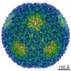 3571C  3573C 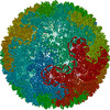 5muuC  5muwC M: map data used to model this data C: citing same article ( |
|---|---|
| Similar structure data |


-Links
 PDBj
PDBj- Assembly
| Deposited unit | 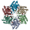
|
|---|---|
| 1 |
|
-Components
| #1: Protein | Mass: 32678.740 Da / Num. of mol.: 6 / Fragment: RESIDUES 1-309 Source method: isolated from a genetically manipulated source Source: (gene. exp.) Pseudomonas phage phi6 (bacteriophage) / Gene: P4 / Production host:  Escherichia coli (E. coli) Escherichia coli (E. coli)References: UniProt: P11125, nucleoside-triphosphate phosphatase #2: Chemical | ChemComp-CA /   Mass: 40.078 Da / Num. of mol.: 6 / Source method: obtained synthetically / Formula: Ca Mass: 40.078 Da / Num. of mol.: 6 / Source method: obtained synthetically / Formula: Ca#3: Chemical | ChemComp-ADP / Adenosine diphosphate  Mass: 427.201 Da / Num. of mol.: 6 / Source method: obtained synthetically / Formula: C10H15N5O10P2 / Comment: ADP, energy-carrying molecule*YM Mass: 427.201 Da / Num. of mol.: 6 / Source method: obtained synthetically / Formula: C10H15N5O10P2 / Comment: ADP, energy-carrying molecule*YM |
|---|
-Experimental details
-Experiment
| Experiment | Method: ELECTRON MICROSCOPY |
|---|---|
| EM experiment | Aggregation state: PARTICLE / 3D reconstruction method: single particle reconstruction |
- Sample preparation
| Component | Name: Pseudomonas phage phi6Pseudomonas virus phi6 / Type: VIRUS / Entity ID: #1 / Source: NATURAL |
|---|---|
| Source (natural) | Organism: Pseudomonas phage phi6 (bacteriophage) |
| Details of virus | Empty: NO / Enveloped: YES / Isolate: SPECIES / Type: VIRION |
| Natural host | Organism: Pseudomonas syringae |
| Buffer solution | pH: 7.2 |
| Specimen | Conc.: 3 mg/ml / Embedding applied: NO / Shadowing applied: NO / Staining applied: NO / Vitrification applied: YES |
| Specimen support | Grid material: COPPER / Grid type: C-flat |
| Vitrification | Cryogen name: ETHANE |
- Electron microscopy imaging
| Experimental equipment |  Model: Tecnai Polara / Image courtesy: FEI Company |
|---|---|
| Microscopy | Model: FEI POLARA 300 |
| Electron gun | Electron source: FIELD EMISSION GUN / Accelerating voltage: 300 kV / Illumination mode: FLOOD BEAM |
| Electron lens | Mode: BRIGHT FIELDBright-field microscopy / Calibrated magnification: 37037 X / Calibrated defocus min: 300 nm / Calibrated defocus max: 3000 nm / Cs: 2 mm / C2 aperture diameter: 50 µm / Alignment procedure: COMA FREE |
| Specimen holder | Cryogen: NITROGEN / Specimen holder model: OTHER / Temperature (max): 120 K / Temperature (min): 80 K |
| Image recording | Average exposure time: 0.2 sec. / Electron dose: 0.7 e/Å2 / Detector mode: COUNTING / Film or detector model: GATAN K2 QUANTUM (4k x 4k) / Num. of real images: 900 |
| EM imaging optics | Energyfilter name: GIF Quantum LS / Energyfilter upper: 20 eV / Energyfilter lower: 0 eV |
| Image scans | Sampling size: 5 µm / Width: 3710 / Height: 3710 / Movie frames/image: 22 / Used frames/image: 1-22 |
- Processing
| EM software |
| |||||||||||||||||||||||||||||||||||||||||||||
|---|---|---|---|---|---|---|---|---|---|---|---|---|---|---|---|---|---|---|---|---|---|---|---|---|---|---|---|---|---|---|---|---|---|---|---|---|---|---|---|---|---|---|---|---|---|---|
| CTF correction | Type: PHASE FLIPPING AND AMPLITUDE CORRECTION | |||||||||||||||||||||||||||||||||||||||||||||
| Particle selection | Num. of particles selected: 159492 | |||||||||||||||||||||||||||||||||||||||||||||
| Symmetry | Point symmetry: C6 (6 fold cyclic) | |||||||||||||||||||||||||||||||||||||||||||||
| 3D reconstruction | Resolution: 9.1 Å / Resolution method: FSC 0.143 CUT-OFF / Num. of particles: 56448 / Algorithm: FOURIER SPACE / Symmetry type: POINT | |||||||||||||||||||||||||||||||||||||||||||||
| Atomic model building | Protocol: RIGID BODY FIT / Space: REAL / Target criteria: Cross-correlation coefficient |
