Movie
Movie Controller


+ Open data

- Basic information
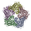
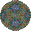
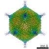
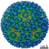
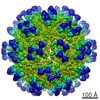
| Entry | Database: PDB / ID: 5muu | ||||||
|---|---|---|---|---|---|---|---|
| Title | dsRNA bacteriophage phi6 nucleocapsid | ||||||
 Components Components |
| ||||||
 Keywords Keywords |  VIRUS / icosahedral virus capsid shell VIRUS / icosahedral virus capsid shell | ||||||
| Function / homology |  Function and homology information Function and homology informationT=13 icosahedral viral capsid / T=2 icosahedral viral capsid / viral procapsid / viral genome packaging / viral inner capsid / viral outer capsid / virion component => GO:0044423 / ribonucleoside triphosphate phosphatase activity / viral capsid / nucleoside-triphosphate phosphatase ...T=13 icosahedral viral capsid / T=2 icosahedral viral capsid / viral procapsid / viral genome packaging / viral inner capsid / viral outer capsid / virion component => GO:0044423 / ribonucleoside triphosphate phosphatase activity / viral capsid / nucleoside-triphosphate phosphatase / viral nucleocapsid / RNA binding / ATP binding / identical protein bindingSimilarity search - Function | ||||||
| Biological species |  Pseudomonas phage phi6 (bacteriophage) Pseudomonas phage phi6 (bacteriophage) | ||||||
| Method | ELECTRON MICROSCOPY / single particle reconstruction / cryo EM / Resolution: 4 Å | ||||||
 Authors Authors | Sun, Z. / El Omari, K. / Sun, X. / Ilca, S.L. / Kotecha, A. / Stuart, D.I. / Poranen, M.M. / Huiskonen, J.T. | ||||||
 Citation Citation | Journal: Nat Commun / Year: 2017 Title: Double-stranded RNA virus outer shell assembly by bona fide domain-swapping. Authors: Zhaoyang Sun / Kamel El Omari / Xiaoyu Sun / Serban L Ilca / Abhay Kotecha / David I Stuart / Minna M Poranen / Juha T Huiskonen /   Abstract: Correct outer protein shell assembly is a prerequisite for virion infectivity in many multi-shelled dsRNA viruses. In the prototypic dsRNA bacteriophage φ6, the assembly reaction is promoted by ...Correct outer protein shell assembly is a prerequisite for virion infectivity in many multi-shelled dsRNA viruses. In the prototypic dsRNA bacteriophage φ6, the assembly reaction is promoted by calcium ions but its biomechanics remain poorly understood. Here, we describe the near-atomic resolution structure of the φ6 double-shelled particle. The outer T=13 shell protein P8 consists of two alpha-helical domains joined by a linker, which allows the trimer to adopt either a closed or an open conformation. The trimers in an open conformation swap domains with each other. Our observations allow us to propose a mechanistic model for calcium concentration regulated outer shell assembly. Furthermore, the structure provides a prime exemplar of bona fide domain-swapping. This leads us to extend the theory of domain-swapping from the level of monomeric subunits and multimers to closed spherical shells, and to hypothesize a mechanism by which closed protein shells may arise in evolution. | ||||||
| History |
|
- Structure visualization
| Movie |
Movie viewer |
|---|---|
| Structure viewer | Molecule: MolmilJmol/JSmol |
- Downloads & links
-Download
| PDBx/mmCIF format | 5muu.cif.gz | 545.2 KB | Display | PDBx/mmCIF format |
|---|---|---|---|---|
| PDB format | pdb5muu.ent.gz | 464.2 KB | Display | PDB format |
| PDBx/mmJSON format | 5muu.json.gz | Tree view | PDBx/mmJSON format | |
| Others |  Other downloads Other downloads |
-Validation report
| Arichive directory | https://data.pdbj.org/pub/pdb/validation_reports/mu/5muuftp://data.pdbj.org/pub/pdb/validation_reports/mu/5muu | HTTPS FTP |
|---|
-Related structure data
| Related structure data | 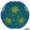 3571MC  3572C  3573C  5muvC  5muwC M: map data used to model this data C: citing same article ( |
|---|---|
| Similar structure data |

-Links
 PDBj
PDBj
- Assembly
| Deposited unit | 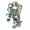
|
|---|---|
| 1 | x 60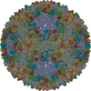
|
| 2 |
|
| 3 | x 5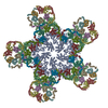
|
| 4 | x 6
|
| 5 | 
|
| Symmetry | Point symmetry: (Schoenflies symbol: I (icosahedral)) |
-Components
| #1: Protein | Mass: 85080.711 Da / Num. of mol.: 2 / Source method: isolated from a natural source / Source: (natural) Pseudomonas phage phi6 (bacteriophage) / References: UniProt: P11126#2: Protein | | Mass: 35198.426 Da / Num. of mol.: 1 / Source method: isolated from a natural source / Source: (natural) Pseudomonas phage phi6 (bacteriophage)References: UniProt: P11125, nucleoside-triphosphate phosphatase #3: Protein | Mass: 16018.418 Da / Num. of mol.: 10 / Source method: isolated from a natural source / Source: (natural) Pseudomonas phage phi6 (bacteriophage) / References: UniProt: P07579 |
|---|
-Experimental details
-Experiment
| Experiment | Method: ELECTRON MICROSCOPY |
|---|---|
| EM experiment | Aggregation state: PARTICLE / 3D reconstruction method: single particle reconstruction |
| Crystal symmetry | ∠γ: 90 ° / A: 1 Å / B: 1 Å / C: 1 Å / Space group name H-M: P1 |
- Sample preparation
| Component | Name: Pseudomonas phage phi6Pseudomonas virus phi6 / Type: VIRUS Details: The viral envelope was removed by Triton X-114 extraction Entity ID: all / Source: NATURAL | |||||||||||||||
|---|---|---|---|---|---|---|---|---|---|---|---|---|---|---|---|---|
| Molecular weight | Experimental value: NO | |||||||||||||||
| Source (natural) | Organism: Pseudomonas phage phi6 (bacteriophage) | |||||||||||||||
| Details of virus | Empty: NO / Enveloped: YES / Isolate: SPECIES / Type: VIRION | |||||||||||||||
| Natural host | Organism: Pseudomonas syringae / Strain: pv.phaseolicola HB10Y | |||||||||||||||
| Virus shell |
| |||||||||||||||
| Buffer solution | pH: 7.2 | |||||||||||||||
| Specimen | Conc.: 3 mg/ml / Embedding applied: NO / Shadowing applied: NO / Staining applied: NO / Vitrification applied: YES | |||||||||||||||
| Specimen support | Grid material: COPPER / Grid type: C-flat | |||||||||||||||
| Vitrification | Instrument: FEI VITROBOT MARK III / Cryogen name: ETHANE |
- Electron microscopy imaging
| Experimental equipment |  Model: Tecnai Polara / Image courtesy: FEI Company |
|---|---|
| Microscopy | Model: FEI POLARA 300 |
| Electron gun | Electron source: FIELD EMISSION GUN / Accelerating voltage: 300 kV / Illumination mode: FLOOD BEAM |
| Electron lens | Mode: BRIGHT FIELDBright-field microscopy / Nominal magnification: 160000 X / Calibrated magnification: 37037 X / Nominal defocus max: 300 nm / Calibrated defocus max: 3000 nm / Cs: 2 mm / C2 aperture diameter: 50 µm / Alignment procedure: COMA FREE |
| Specimen holder | Cryogen: NITROGEN / Specimen holder model: OTHER / Temperature (max): 120 K / Temperature (min): 80 K |
| Image recording | Average exposure time: 0.2 sec. / Electron dose: 0.73 e/Å2 / Detector mode: COUNTING / Film or detector model: GATAN K2 SUMMIT (4k x 4k) / Num. of real images: 900 |
| EM imaging optics | Energyfilter name: GIF Quantum LS / Energyfilter upper: 20 eV / Energyfilter lower: 0 eV |
| Image scans | Sampling size: 5 µm / Width: 3710 / Height: 3710 / Movie frames/image: 22 / Used frames/image: 1-22 |
- Processing
| Software | Name: PHENIX / Version: dev_2044: / Classification: refinement | ||||||||||||||||||||||||||||||||||||||||||||||||||||||||||||||||||||||||||||||||||||||||||||||||||||||||||||
|---|---|---|---|---|---|---|---|---|---|---|---|---|---|---|---|---|---|---|---|---|---|---|---|---|---|---|---|---|---|---|---|---|---|---|---|---|---|---|---|---|---|---|---|---|---|---|---|---|---|---|---|---|---|---|---|---|---|---|---|---|---|---|---|---|---|---|---|---|---|---|---|---|---|---|---|---|---|---|---|---|---|---|---|---|---|---|---|---|---|---|---|---|---|---|---|---|---|---|---|---|---|---|---|---|---|---|---|---|---|
| EM software |
| ||||||||||||||||||||||||||||||||||||||||||||||||||||||||||||||||||||||||||||||||||||||||||||||||||||||||||||
| Crystal symmetry | ∠γ: 90 ° / A: 1 Å / B: 1 Å / C: 1 Å / Space group name H-M: P1 | ||||||||||||||||||||||||||||||||||||||||||||||||||||||||||||||||||||||||||||||||||||||||||||||||||||||||||||
| CTF correction | Type: PHASE FLIPPING AND AMPLITUDE CORRECTION | ||||||||||||||||||||||||||||||||||||||||||||||||||||||||||||||||||||||||||||||||||||||||||||||||||||||||||||
| Particle selection | Num. of particles selected: 16466 | ||||||||||||||||||||||||||||||||||||||||||||||||||||||||||||||||||||||||||||||||||||||||||||||||||||||||||||
| Symmetry | Point symmetry: I (icosahedral) | ||||||||||||||||||||||||||||||||||||||||||||||||||||||||||||||||||||||||||||||||||||||||||||||||||||||||||||
| 3D reconstruction | Resolution: 4 Å / Resolution method: FSC 0.143 CUT-OFF / Num. of particles: 13291 / Algorithm: FOURIER SPACE / Symmetry type: POINT | ||||||||||||||||||||||||||||||||||||||||||||||||||||||||||||||||||||||||||||||||||||||||||||||||||||||||||||
| Atomic model building |
| ||||||||||||||||||||||||||||||||||||||||||||||||||||||||||||||||||||||||||||||||||||||||||||||||||||||||||||
| Atomic model building |
| ||||||||||||||||||||||||||||||||||||||||||||||||||||||||||||||||||||||||||||||||||||||||||||||||||||||||||||
| Refine LS restraints |
|