 Movie
Movie Controller

[English] 日本語
 Yorodumi
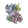
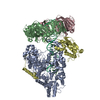
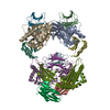
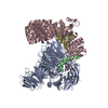
- PDB-5m1s: Cryo-EM structure of the E. coli replicative DNA polymerase-clamp... -
+ Open data

- Basic information
| Entry | Database: PDB / ID: 5m1s | ||||||
|---|---|---|---|---|---|---|---|
| Title | Cryo-EM structure of the E. coli replicative DNA polymerase-clamp-exonuclase-theta complex bound to DNA in the editing mode | ||||||
 Components Components |
| ||||||
 Keywords Keywords |  DNA BINDING PROTEIN / DNA editing Proofreading Exonuclease Polymerase DNA BINDING PROTEIN / DNA editing Proofreading Exonuclease Polymerase | ||||||
| Function / homology |  Function and homology informationDNA polymerase III, core complex / Hda-beta clamp complex / bacterial-type DNA replication / replication inhibiting complex / DNA replication proofreading / DNA polymerase III complex / lagging strand elongation / replisome / regulation of DNA-templated DNA replication initiation / DNA strand elongation involved in DNA replication ...DNA polymerase III, core complex / Hda-beta clamp complex / bacterial-type DNA replication / replication inhibiting complex / DNA replication proofreading / DNA polymerase III complex / lagging strand elongation / replisome / regulation of DNA-templated DNA replication initiation / DNA strand elongation involved in DNA replication / exonuclease activity / leading strand elongation / error-prone translesion synthesis / negative regulation of DNA-templated DNA replication initiation / 3'-5' exonuclease activity / DNA-templated DNA replication / DNA-directed DNA polymerase / DNA-directed DNA polymerase activity / DNA damage response / protein homodimerization activity / DNA binding / identical protein binding / metal ion binding / cytosol / cytoplasm Function and homology informationDNA polymerase III, core complex / Hda-beta clamp complex / bacterial-type DNA replication / replication inhibiting complex / DNA replication proofreading / DNA polymerase III complex / lagging strand elongation / replisome / regulation of DNA-templated DNA replication initiation / DNA strand elongation involved in DNA replication ...DNA polymerase III, core complex / Hda-beta clamp complex / bacterial-type DNA replication / replication inhibiting complex / DNA replication proofreading / DNA polymerase III complex / lagging strand elongation / replisome / regulation of DNA-templated DNA replication initiation / DNA strand elongation involved in DNA replication / exonuclease activity / leading strand elongation / error-prone translesion synthesis / negative regulation of DNA-templated DNA replication initiation / 3'-5' exonuclease activity / DNA-templated DNA replication / DNA-directed DNA polymerase / DNA-directed DNA polymerase activity / DNA damage response / protein homodimerization activity / DNA binding / identical protein binding / metal ion binding / cytosol / cytoplasmSimilarity search - Function | ||||||
| Biological species |  Escherichia coli K12 (bacteria) Escherichia coli K12 (bacteria)synthetic construct (others) | ||||||
| Method | ELECTRON MICROSCOPY / single particle reconstruction / cryo EM / Resolution: 6.7 Å | ||||||
 Authors Authors | Fernandez-Leiro, R. / Conrad, J. / Scheres, S.H.W. / Lamers, M.H. | ||||||
 Citation Citation | Journal: Nat Struct Mol Biol / Year: 2017 Title: Self-correcting mismatches during high-fidelity DNA replication. Authors: Rafael Fernandez-Leiro / Julian Conrad / Ji-Chun Yang / Stefan M V Freund / Sjors H W Scheres / Meindert H Lamers /  Abstract: Faithful DNA replication is essential to all forms of life and depends on the action of 3'-5' exonucleases that remove misincorporated nucleotides from the newly synthesized strand. However, how the ...Faithful DNA replication is essential to all forms of life and depends on the action of 3'-5' exonucleases that remove misincorporated nucleotides from the newly synthesized strand. However, how the DNA is transferred from the polymerase to the exonuclease active site is not known. Here we present the cryo-EM structure of the editing mode of the catalytic core of the Escherichia coli replisome, revealing a dramatic distortion of the DNA whereby the polymerase thumb domain acts as a wedge that separates the two DNA strands. Importantly, NMR analysis of the DNA substrate shows that the presence of a mismatch increases the fraying of the DNA, thus enabling it to reach the exonuclease active site. Therefore the mismatch corrects itself, whereas the exonuclease subunit plays a passive role. Hence, our work provides unique insights into high-fidelity replication and establishes a new paradigm for the correction of misincorporated nucleotides. | ||||||
| History |
|
- Structure visualization
| Movie |
Movie viewer |
|---|---|
| Structure viewer | Molecule: MolmilJmol/JSmol |
- Downloads & links
-Download
| PDBx/mmCIF format | 5m1s.cif.gz | 351.2 KB | Display | PDBx/mmCIF format |
|---|---|---|---|---|
| PDB format | pdb5m1s.ent.gz | 281.5 KB | Display | PDB format |
| PDBx/mmJSON format | 5m1s.json.gz | Tree view | PDBx/mmJSON format | |
| Others |  Other downloads Other downloads |
-Validation report
| Arichive directory | https://data.pdbj.org/pub/pdb/validation_reports/m1/5m1sftp://data.pdbj.org/pub/pdb/validation_reports/m1/5m1s | HTTPS FTP |
|---|
-Related structure data
| Related structure data |  4141MC 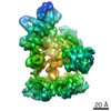 4142C M: map data used to model this data C: citing same article ( |
|---|---|
| Similar structure data |


-Links
 PDBj
PDBj


- Assembly
| Deposited unit | 
|
|---|---|
| 1 |
|
-Components
-DNA polymerase III subunit ... , 4 types, 5 molecules ABCDF
| #1: Protein | DNA polymerase III holoenzyme Mass: 103554.422 Da / Num. of mol.: 1 / Mutation: A921L, M923L Source method: isolated from a genetically manipulated source Source: (gene. exp.) Escherichia coli K12 (bacteria) / Gene: dnaE, polC, b0184, JW0179 / Production host: Escherichia coli BL21(DE3) (bacteria) / References: UniProt: P10443, DNA-directed DNA polymerase | ||||
|---|---|---|---|---|---|
| #2: Protein | DNA polymerase III holoenzyme / Beta sliding clamp / Beta clamp Mass: 40630.508 Da / Num. of mol.: 2 Source method: isolated from a genetically manipulated source Source: (gene. exp.) Escherichia coli K12 (bacteria) / Gene: dnaN, b3701, JW3678 / Production host: Escherichia coli BL21(DE3) (bacteria) / References: UniProt: P0A988, DNA-directed DNA polymerase#3: Protein | | DNA polymerase III holoenzymeMass: 27118.984 Da / Num. of mol.: 1 / Mutation: T183L M185L A186P F187L Source method: isolated from a genetically manipulated source Source: (gene. exp.) Escherichia coli K12 (bacteria) / Gene: dnaQ, mutD, b0215, JW0205 / Production host: Escherichia coli BL21(DE3) (bacteria) / References: UniProt: P03007, DNA-directed DNA polymerase#6: Protein | | DNA polymerase III holoenzymeMass: 6503.385 Da / Num. of mol.: 1 Source method: isolated from a genetically manipulated source Source: (gene. exp.) Escherichia coli K12 (bacteria) / Gene: holE, b1842, JW1831 / Production host: Escherichia coli BL21(DE3) (bacteria) / References: UniProt: P0ABS8, DNA-directed DNA polymerase |
-DNA chain , 2 types, 2 molecules PT
| #4: DNA chain | Mass: 5275.448 Da / Num. of mol.: 1 Source method: isolated from a genetically manipulated source Source: (gene. exp.) synthetic construct (others) / Production host: synthetic construct (others) |
|---|---|
| #5: DNA chain | Mass: 6718.339 Da / Num. of mol.: 1 Source method: isolated from a genetically manipulated source Source: (gene. exp.) synthetic construct (others) / Production host: synthetic construct (others) |
-Experimental details
-Experiment
| Experiment | Method: ELECTRON MICROSCOPY |
|---|---|
| EM experiment | Aggregation state: PARTICLE / 3D reconstruction method: single particle reconstruction |
- Sample preparation
| Component |
| ||||||||||||||||||||||||||||
|---|---|---|---|---|---|---|---|---|---|---|---|---|---|---|---|---|---|---|---|---|---|---|---|---|---|---|---|---|---|
| Molecular weight |
| ||||||||||||||||||||||||||||
| Source (natural) |
| ||||||||||||||||||||||||||||
| Source (recombinant) |
| ||||||||||||||||||||||||||||
| Buffer solution | pH: 7.5 | ||||||||||||||||||||||||||||
| Buffer component |
| ||||||||||||||||||||||||||||
| Specimen | Conc.: 0.25 mg/ml / Embedding applied: NO / Shadowing applied: NO / Staining applied: NO / Vitrification applied: YES Details: Sample was run over a gel filtration column prior to vitrification | ||||||||||||||||||||||||||||
| Specimen support | Grid material: COPPER / Grid mesh size: 300 divisions/in. / Grid type: Quantifoil R1.2/1.3 | ||||||||||||||||||||||||||||
| Vitrification | Instrument: FEI VITROBOT MARK IV / Cryogen name: ETHANE / Humidity: 100 % / Chamber temperature: 277 K Details: Prior to sample preparation 0.1 volumes of 0.05% Tween 20 were added to the sample 3 microliters were pipetted onto the grid and blotted for 4 seconds |
- Electron microscopy imaging
| Experimental equipment |  Model: Titan Krios / Image courtesy: FEI Company |
|---|---|
| Microscopy | Model: FEI TITAN KRIOS |
| Electron gun | Electron source: FIELD EMISSION GUN / Accelerating voltage: 300 kV / Illumination mode: FLOOD BEAM |
| Electron lens | Mode: BRIGHT FIELDBright-field microscopy / Nominal magnification: 64000 X / Calibrated magnification: 79545 X / Nominal defocus max: 3500 nm / Nominal defocus min: 1800 nm / Cs: 2.7 mm / C2 aperture diameter: 50 µm / Alignment procedure: COMA FREE |
| Specimen holder | Cryogen: NITROGEN / Specimen holder model: FEI TITAN KRIOS AUTOGRID HOLDER / Temperature (max): 80 K / Temperature (min): 80 K |
| Image recording | Average exposure time: 25 sec. / Electron dose: 2 e/Å2 / Detector mode: COUNTING / Film or detector model: GATAN K2 SUMMIT (4k x 4k) / Num. of grids imaged: 3 / Num. of real images: 1157 |
| EM imaging optics | Energyfilter name: GIF Quantum / Energyfilter upper: 20 eV / Energyfilter lower: 0 eV |
| Image scans | Width: 3710 / Height: 3710 / Movie frames/image: 20 / Used frames/image: 1-20 |
- Processing
| EM software |
| ||||||||||||||||||||||||||||||||||||||||||||||||||||||||||||
|---|---|---|---|---|---|---|---|---|---|---|---|---|---|---|---|---|---|---|---|---|---|---|---|---|---|---|---|---|---|---|---|---|---|---|---|---|---|---|---|---|---|---|---|---|---|---|---|---|---|---|---|---|---|---|---|---|---|---|---|---|---|
| CTF correction | Type: NONE | ||||||||||||||||||||||||||||||||||||||||||||||||||||||||||||
| Particle selection | Num. of particles selected: 150000 | ||||||||||||||||||||||||||||||||||||||||||||||||||||||||||||
| Symmetry | Point symmetry: C1 (asymmetric) | ||||||||||||||||||||||||||||||||||||||||||||||||||||||||||||
| 3D reconstruction | Resolution: 6.7 Å / Resolution method: FSC 0.143 CUT-OFF / Num. of particles: 15616 / Algorithm: FOURIER SPACE / Symmetry type: POINT | ||||||||||||||||||||||||||||||||||||||||||||||||||||||||||||
| Atomic model building | Protocol: OTHER Details: The cryo-EM structure of the PolIIIalpha-clamp-exonuclease complex in the polymerase mode (PDB code: 5FKW) was used as a starting model, and the NMR structure of theta bound to the ...Details: The cryo-EM structure of the PolIIIalpha-clamp-exonuclease complex in the polymerase mode (PDB code: 5FKW) was used as a starting model, and the NMR structure of theta bound to the exonuclease catalytic domain (PDB code: 2XY8) was used to place theta into the cryo-EM map. The model was manually adjusted in Coot and geometry of the protein optimized in Refmac5 using DNA-specific restraints generated in LibG |
