 Movie
Movie Controller

[English] 日本語
 Yorodumi
- PDB-4bom: Structure of herpesvirus fusion glycoprotein B-bilayer complex re... -
+ Open data

- Basic information
| Entry | Database: PDB / ID: 4bom | ||||||
|---|---|---|---|---|---|---|---|
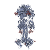
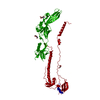
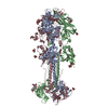
| Title | Structure of herpesvirus fusion glycoprotein B-bilayer complex revealing the protein-membrane and lateral protein-protein interaction | ||||||
 Components Components | ENVELOPE GLYCOPROTEIN B | ||||||
 Keywords Keywords |  VIRAL PROTEIN / MEMBRANE PROXIMAL REGION / PROTEIN COAT / PSEUDO-ATOMIC VIRUS-HOST INTERACTION VIRAL PROTEIN / MEMBRANE PROXIMAL REGION / PROTEIN COAT / PSEUDO-ATOMIC VIRUS-HOST INTERACTION | ||||||
| Function / homology |  Function and homology information Function and homology informationhost cell Golgi membrane / host cell endosome membrane / symbiont entry into host cell / viral envelope / virion attachment to host cell / host cell plasma membrane / virion membrane / membrane / identical protein bindingSimilarity search - Function | ||||||
| Biological species |   HUMAN HERPESVIRUS 1 (Herpes simplex virus type 1) HUMAN HERPESVIRUS 1 (Herpes simplex virus type 1) | ||||||
| Method | ELECTRON MICROSCOPY / electron tomography / cryo EM / Resolution: 27 Å | ||||||
 Authors Authors | Maurer, U.E. / Zeev-Ben-Mordehai, Z. / Pandurangan, A.P. / Cairns, T.M. / Hannah, B.P. / Whitbeck, J.C. / Eisenberg, R.J. / Cohen, G.H. / Topf, M. / Huiskonen, J.T. / Grunewald, K. | ||||||
 Citation Citation | Journal: Structure / Year: 2013 Title: The structure of herpesvirus fusion glycoprotein B-bilayer complex reveals the protein-membrane and lateral protein-protein interaction. Authors: Ulrike E Maurer / Tzviya Zeev-Ben-Mordehai / Arun Prasad Pandurangan / Tina M Cairns / Brian P Hannah / J Charles Whitbeck / Roselyn J Eisenberg / Gary H Cohen / Maya Topf / Juha T Huiskonen / Kay Grünewald /  Abstract: Glycoprotein B (gB) is a key component of the complex herpesvirus fusion machinery. We studied membrane interaction of two gB ectodomain forms and present an electron cryotomography structure of the ...Glycoprotein B (gB) is a key component of the complex herpesvirus fusion machinery. We studied membrane interaction of two gB ectodomain forms and present an electron cryotomography structure of the gB-bilayer complex. The two forms differed in presence or absence of the membrane proximal region (MPR) but showed an overall similar trimeric shape. The presence of the MPR impeded interaction with liposomes. In contrast, the MPR-lacking form interacted efficiently with liposomes. Lateral interaction resulted in coat formation on the membranes. The structure revealed that interaction of gB with membranes was mediated by the fusion loops and limited to the outer membrane leaflet. The observed intrinsic propensity of gB to cluster on membranes indicates an additional role of gB in driving the fusion process forward beyond the transient fusion pore opening and subsequently leading to fusion pore expansion. | ||||||
| History |
| ||||||
| Remark 700 | SHEET DETERMINATION METHOD: DSSP THE SHEETS PRESENTED AS "AC" IN EACH CHAIN ON SHEET RECORDS BELOW ... SHEET DETERMINATION METHOD: DSSP THE SHEETS PRESENTED AS "AC" IN EACH CHAIN ON SHEET RECORDS BELOW IS ACTUALLY AN 6-STRANDED BARREL THIS IS REPRESENTED BY A 7-STRANDED SHEET IN WHICH THE FIRST AND LAST STRANDS ARE IDENTICAL. THE SHEETS PRESENTED AS "BC" IN EACH CHAIN ON SHEET RECORDS BELOW IS ACTUALLY AN 6-STRANDED BARREL THIS IS REPRESENTED BY A 7-STRANDED SHEET IN WHICH THE FIRST AND LAST STRANDS ARE IDENTICAL. THE SHEETS PRESENTED AS "CB" IN EACH CHAIN ON SHEET RECORDS BELOW IS ACTUALLY AN 6-STRANDED BARREL THIS IS REPRESENTED BY A 7-STRANDED SHEET IN WHICH THE FIRST AND LAST STRANDS ARE IDENTICAL. |
- Structure visualization
| Movie |
Movie viewer |
|---|---|
| Structure viewer | Molecule: MolmilJmol/JSmol |
- Downloads & links
-Download
| PDBx/mmCIF format | 4bom.cif.gz | 325 KB | Display | PDBx/mmCIF format |
|---|---|---|---|---|
| PDB format | pdb4bom.ent.gz | 274.4 KB | Display | PDB format |
| PDBx/mmJSON format | 4bom.json.gz | Tree view | PDBx/mmJSON format | |
| Others |  Other downloads Other downloads |
-Validation report
| Arichive directory | https://data.pdbj.org/pub/pdb/validation_reports/bo/4bomftp://data.pdbj.org/pub/pdb/validation_reports/bo/4bom | HTTPS FTP |
|---|
-Related structure data
| Related structure data |  2380MC 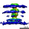 2379C M: map data used to model this data C: citing same article ( |
|---|---|
| Similar structure data |


-Links
 PDBj
PDBj- Assembly
| Deposited unit | 
|
|---|---|
| 1 |
|
-Components
| #1: Protein | Mass: 71125.688 Da / Num. of mol.: 3 / Fragment: GLYCOPROTEIN B ECTODOMAIN, RESIDUES 103-724 Source method: isolated from a genetically manipulated source Details: LIPOSOMES CONSISTING OF PHOSPHATIDYLCHOLINE AND CHOLESTEROL AT 1.7\:1 MOLAR RATIO WERE INCUBATED WITH GB AT PH 5.5 AT 37C FOR ONE HOUR Source: (gene. exp.) HUMAN HERPESVIRUS 1 (Herpes simplex virus type 1)Strain: KOS / Plasmid: PCW289 / Cell line (production host): Sf9 / Production host: Spodoptera frugiperda / References: UniProt: P06437 |
|---|
-Experimental details
-Experiment
| Experiment | Method: ELECTRON MICROSCOPY |
|---|---|
| EM experiment | Aggregation state: PARTICLE / 3D reconstruction method: electron tomography |
- Sample preparation
| Component | Name: HUMAN HERPESVIRUS 1 ENVELOPED GLYCOPROTEIN GB ECTODOMAIN LACKING THE MEMBRANE PROXIMAL REGION BOUND TO LIPOSOMES Type: COMPLEX / Source: RECOMBINANT |
|---|---|
| Source (natural) | Organism: HUMAN HERPESVIRUS 1 (Herpes simplex virus type 1) |
| Source (recombinant) | Organism:   Spodoptera frugiperda (fall armyworm) Spodoptera frugiperda (fall armyworm) |
| Buffer solution | Name: PBS WITH SODIUM CITRATE / pH: 5.5 / Details: PBS WITH SODIUM CITRATE |
| Specimen | Conc.: 1 mg/ml / Embedding applied: NO / Shadowing applied: NO / Staining applied: NO / Vitrification applied: YES |
| Specimen support | Details: HOLEY CARBON |
| Vitrification | Cryogen name: ETHANE-PROPANE / Details: LIQUID NITROGEN PROPANE MIXTURE |
- Electron microscopy imaging
| Experimental equipment |  Model: Tecnai F20 / Image courtesy: FEI Company |
|---|---|
| Microscopy | Model: FEI TECNAI F20 / Date: Jan 17, 2008 |
| Electron gun | Electron source: FIELD EMISSION GUN / Accelerating voltage: 200 kV / Illumination mode: FLOOD BEAM |
| Electron lens | Mode: BRIGHT FIELDBright-field microscopy / Calibrated magnification: 67000 X / Nominal defocus max: 2000 nm / Nominal defocus min: 2000 nm / Cs: 2 mm |
| Specimen holder | Tilt angle max: 60 ° / Tilt angle min: -60 ° |
| Image recording | Electron dose: 100 e/Å2 / Film or detector model: GATAN ULTRASCAN 4000 (4k x 4k) |
| Image scans | Num. digital images: 9 |
- Processing
| EM software |
| ||||||||||||
|---|---|---|---|---|---|---|---|---|---|---|---|---|---|
| CTF correction | Details: LOW PASS FILTER TO THE FIRST ZERO CROSSING OF THE CTF | ||||||||||||
| Symmetry | Point symmetry: C3 (3 fold cyclic) | ||||||||||||
| 3D reconstruction | Method: SUBTOMOGRAM AVERAGING / Resolution: 27 Å / Num. of particles: 786 / Actual pixel size: 4.6 Å Details: THE MISSING FUSION LOOP RESIDUES 261 AND 262 WERE MODELED IN ALL THE THREE CHAINS OF USING MODELLER BASED ON THE GB CRYSTAL STRUCTURE (PDB ID 2GUM) SUBMISSION BASED ON EXPERIMENTAL DATA FROM ...Details: THE MISSING FUSION LOOP RESIDUES 261 AND 262 WERE MODELED IN ALL THE THREE CHAINS OF USING MODELLER BASED ON THE GB CRYSTAL STRUCTURE (PDB ID 2GUM) SUBMISSION BASED ON EXPERIMENTAL DATA FROM EMDB EMD-2380. (DEPOSITION ID: 11670). Symmetry type: POINT | ||||||||||||
| Atomic model building | Protocol: FLEXIBLE FIT / Space: REAL / Details: METHOD--FLEXIBLE REFINEMENT PROTOCOL--FLEXIBLE | ||||||||||||
| Atomic model building | PDB-ID: 3NWF | ||||||||||||
| Refinement | Highest resolution: 27 Å | ||||||||||||
| Refinement step | Cycle: LAST / Highest resolution: 27 Å
|

