 Movie
Movie Controller


+ Open data

- Basic information
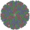
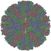
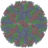
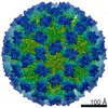
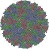
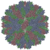
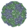
| Entry | Database: PDB / ID: 3zue | ||||||
|---|---|---|---|---|---|---|---|
| Title | Rabbit Hemorrhagic Disease Virus (RHDV)capsid protein | ||||||
 Components Components | CAPSID STRUCTURAL PROTEIN VP60 | ||||||
 Keywords Keywords |  VIRUS / CAGE DESIGN / MOLECULAR SWITCH / PROTEIN ENGINEERING / STRUCTURAL POLYMORPHISM / VIRUS ASSEMBLY VIRUS / CAGE DESIGN / MOLECULAR SWITCH / PROTEIN ENGINEERING / STRUCTURAL POLYMORPHISM / VIRUS ASSEMBLY | ||||||
| Function / homology |  Function and homology informationcalicivirin / ribonucleoside triphosphate phosphatase activity / viral capsid / nucleoside-triphosphate phosphatase / host cell cytoplasm / RNA helicase activity / RNA-directed RNA polymerase / viral RNA genome replication / cysteine-type endopeptidase activity / RNA-dependent RNA polymerase activity ...calicivirin / ribonucleoside triphosphate phosphatase activity / viral capsid / nucleoside-triphosphate phosphatase / host cell cytoplasm / RNA helicase activity / RNA-directed RNA polymerase / viral RNA genome replication / cysteine-type endopeptidase activity / RNA-dependent RNA polymerase activity / DNA-templated transcription / proteolysis / RNA binding / ATP binding Function and homology informationcalicivirin / ribonucleoside triphosphate phosphatase activity / viral capsid / nucleoside-triphosphate phosphatase / host cell cytoplasm / RNA helicase activity / RNA-directed RNA polymerase / viral RNA genome replication / cysteine-type endopeptidase activity / RNA-dependent RNA polymerase activity ...calicivirin / ribonucleoside triphosphate phosphatase activity / viral capsid / nucleoside-triphosphate phosphatase / host cell cytoplasm / RNA helicase activity / RNA-directed RNA polymerase / viral RNA genome replication / cysteine-type endopeptidase activity / RNA-dependent RNA polymerase activity / DNA-templated transcription / proteolysis / RNA binding / ATP bindingSimilarity search - Function | ||||||
| Biological species |  RABBIT HEMORRHAGIC DISEASE VIRUS RABBIT HEMORRHAGIC DISEASE VIRUS | ||||||
| Method | ELECTRON MICROSCOPY / single particle reconstruction / cryo EM / Resolution: 10.3 Å | ||||||
| Model type details | CA ATOMS ONLY, CHAIN A, B, C | ||||||
 Authors Authors | Luque, D. / Gonzalez, J.M. / Gomez-Blanco, J. / Marabini, R. / Chichon, J. / Mena, I. / Angulo, I. / Carrascosa, J.L. / Verdaguer, N. / Trus, B.L. ...Luque, D. / Gonzalez, J.M. / Gomez-Blanco, J. / Marabini, R. / Chichon, J. / Mena, I. / Angulo, I. / Carrascosa, J.L. / Verdaguer, N. / Trus, B.L. / Barcena, J. / Caston, J.R. | ||||||
 Citation Citation | Journal: J Virol / Year: 2012 Title: Epitope insertion at the N-terminal molecular switch of the rabbit hemorrhagic disease virus T = 3 capsid protein leads to larger T = 4 capsids. Authors: Daniel Luque / José M González / Josué Gómez-Blanco / Roberto Marabini / Javier Chichón / Ignacio Mena / Iván Angulo / José L Carrascosa / Nuria Verdaguer / Benes L Trus / Juan ...Authors: Daniel Luque / José M González / Josué Gómez-Blanco / Roberto Marabini / Javier Chichón / Ignacio Mena / Iván Angulo / José L Carrascosa / Nuria Verdaguer / Benes L Trus / Juan Bárcena / José R Castón /  Abstract: Viruses need only one or a few structural capsid proteins to build an infectious particle. This is possible through the extensive use of symmetry and the conformational polymorphism of the structural ...Viruses need only one or a few structural capsid proteins to build an infectious particle. This is possible through the extensive use of symmetry and the conformational polymorphism of the structural proteins. Using virus-like particles (VLP) from rabbit hemorrhagic disease virus (RHDV) as a model, we addressed the basis of calicivirus capsid assembly and their application in vaccine design. The RHDV capsid is based on a T=3 lattice containing 180 identical subunits (VP1). We determined the structure of RHDV VLP to 8.0-Å resolution by three-dimensional cryoelectron microscopy; in addition, we used San Miguel sea lion virus (SMSV) and feline calicivirus (FCV) capsid subunit structures to establish the backbone structure of VP1 by homology modeling and flexible docking analysis. Based on the three-domain VP1 model, several insertion mutants were designed to validate the VP1 pseudoatomic model, and foreign epitopes were placed at the N- or C-terminal end, as well as in an exposed loop on the capsid surface. We selected a set of T and B cell epitopes of various lengths derived from viral and eukaryotic origins. Structural analysis of these chimeric capsids further validates the VP1 model to design new chimeras. Whereas most insertions are well tolerated, VP1 with an FCV capsid protein-neutralizing epitope at the N terminus assembled into mixtures of T=3 and larger T=4 capsids. The calicivirus capsid protein, and perhaps that of many other viruses, thus can encode polymorphism modulators that are not anticipated from the plane sequence, with important implications for understanding virus assembly and evolution. | ||||||
| History |
|
- Structure visualization
| Movie |
Movie viewer |
|---|---|
| Structure viewer | Molecule: MolmilJmol/JSmol |
- Downloads & links
-Download
| PDBx/mmCIF format | 3zue.cif.gz | 61.3 KB | Display | PDBx/mmCIF format |
|---|---|---|---|---|
| PDB format | pdb3zue.ent.gz | 41.7 KB | Display | PDB format |
| PDBx/mmJSON format | 3zue.json.gz | Tree view | PDBx/mmJSON format | |
| Others |  Other downloads Other downloads |
-Validation report
| Arichive directory | https://data.pdbj.org/pub/pdb/validation_reports/zu/3zueftp://data.pdbj.org/pub/pdb/validation_reports/zu/3zue | HTTPS FTP |
|---|
-Related structure data
| Related structure data | 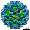 1933MC 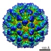 1934C 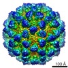 1935C  1936C  1937C 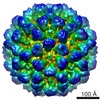 1938C 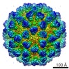 1939C M: map data used to model this data C: citing same article ( |
|---|---|
| Similar structure data |
-Links
 PDBj
PDBj

- Assembly
| Deposited unit | 
|
|---|---|
| 1 | x 60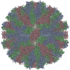
|
| 2 |
|
| 3 | x 5
|
| 4 | x 6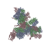
|
| 5 | 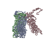
|
| Symmetry | Point symmetry: (Schoenflies symbol: I (icosahedral)) |
-Components
| #1: Protein | Mass: 60264.785 Da / Num. of mol.: 3 Source method: isolated from a genetically manipulated source Source: (gene. exp.) RABBIT HEMORRHAGIC DISEASE VIRUS / Strain: AST89 / Cell line (production host): H5 / Production host:   SPODOPTERA FRUGIPERDA (fall armyworm) / References: UniProt: Q9YND5, UniProt: Q86119*PLUS SPODOPTERA FRUGIPERDA (fall armyworm) / References: UniProt: Q9YND5, UniProt: Q86119*PLUS |
|---|
-Experimental details
-Experiment
| Experiment | Method: ELECTRON MICROSCOPY |
|---|---|
| EM experiment | Aggregation state: PARTICLE / 3D reconstruction method: single particle reconstruction |
- Sample preparation
| Component | Name: WT RABBIT HEMORRHAGIC DISEASE VIRUS VLP / Type: VIRUS |
|---|---|
| Buffer solution | Name: 200 MM NA2HPO4, 200 MM NACL / pH: 6 / Details: 200 MM NA2HPO4, 200 MM NACL |
| Specimen | Embedding applied: NO / Shadowing applied: NO / Staining applied: NO / Vitrification applied: YES |
| Specimen support | Details: HOLEY CARBON |
| Vitrification | Cryogen name: ETHANE / Details: LIQUID ETHANE |
- Electron microscopy imaging
| Experimental equipment |  Model: Tecnai F20 / Image courtesy: FEI Company |
|---|---|
| Microscopy | Model: FEI TECNAI F20 |
| Electron gun | Electron source: FIELD EMISSION GUN / Accelerating voltage: 200 kV / Illumination mode: FLOOD BEAM |
| Electron lens | Mode: BRIGHT FIELDBright-field microscopy / Nominal magnification: 50000 X / Calibrated magnification: 50000 X / Nominal defocus max: 3000 nm / Nominal defocus min: 800 nm / Cs: 2.26 mm |
| Image recording | Electron dose: 10 e/Å2 / Film or detector model: KODAK SO-163 FILM |
| Image scans | Num. digital images: 116 |
- Processing
| EM software | Name: Xmipp / Category: 3D reconstruction | ||||||||||||
|---|---|---|---|---|---|---|---|---|---|---|---|---|---|
| CTF correction | Details: PHASE FLIPPING AND AMPLITUDE DECAY | ||||||||||||
| Symmetry | Point symmetry: I (icosahedral) | ||||||||||||
| 3D reconstruction | Method: PROJECTION MATCHING / Resolution: 10.3 Å / Num. of particles: 5011 / Nominal pixel size: 2.8 Å / Actual pixel size: 2.8 Å Details: SUBMISSION BASED ON EXPERIMENTAL DATA FROM EMDB EMD-1933 (DEPOSITION ID: 10153). Symmetry type: POINT | ||||||||||||
| Atomic model building | Protocol: OTHER / Space: REAL / Details: METHOD--CORRELATION | ||||||||||||
| Refinement | Highest resolution: 10.3 Å | ||||||||||||
| Refinement step | Cycle: LAST / Highest resolution: 10.3 Å
|
