 Movie
Movie Controller

[English] 日本語
 Yorodumi
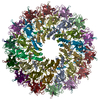
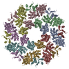
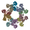
- PDB-3zbi: Fitting result in the O-layer of the subnanometer structure of th... -
+ Open data

- Basic information
| Entry | Database: PDB / ID: 3zbi | ||||||
|---|---|---|---|---|---|---|---|
| Title | Fitting result in the O-layer of the subnanometer structure of the bacterial pKM101 type IV secretion system core complex digested with elastase | ||||||
 Components Components |
| ||||||
 Keywords Keywords |  CELL ADHESION / BACTERIAL SECRETION CELL ADHESION / BACTERIAL SECRETION | ||||||
| Function / homology |  Function and homology information Function and homology information | ||||||
| Biological species |  ESCHERICHIA COLI (E. coli) ESCHERICHIA COLI (E. coli) | ||||||
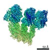
| Method | ELECTRON MICROSCOPY / single particle reconstruction / cryo EM / Resolution: 8.5 Å | ||||||
 Authors Authors | Rivera-Calzada, A. / Fronzes, R. / Savva, C.G. / Chandran, V. / Lian, P.W. / Laeremans, T. / Pardon, E. / Steyaert, J. / Remaut, H. / Waksman, G. / Orlova, E.V. | ||||||
 Citation Citation | Journal: EMBO J / Year: 2013 Title: Structure of a bacterial type IV secretion core complex at subnanometre resolution. Authors: Angel Rivera-Calzada / Rémi Fronzes / Christos G Savva / Vidya Chandran / Pei W Lian / Toon Laeremans / Els Pardon / Jan Steyaert / Han Remaut / Gabriel Waksman / Elena V Orlova /  Abstract: Type IV secretion (T4S) systems are able to transport DNAs and/or proteins through the membranes of bacteria. They form large multiprotein complexes consisting of 12 proteins termed VirB1-11 and ...Type IV secretion (T4S) systems are able to transport DNAs and/or proteins through the membranes of bacteria. They form large multiprotein complexes consisting of 12 proteins termed VirB1-11 and VirD4. VirB7, 9 and 10 assemble into a 1.07 MegaDalton membrane-spanning core complex (CC), around which all other components assemble. This complex is made of two parts, the O-layer inserted in the outer membrane and the I-layer inserted in the inner membrane. While the structure of the O-layer has been solved by X-ray crystallography, there is no detailed structural information on the I-layer. Using high-resolution cryo-electron microscopy and molecular modelling combined with biochemical approaches, we determined the I-layer structure and located its various components in the electron density. Our results provide new structural insights on the CC, from which the essential features of T4S system mechanisms can be derived. #1: Journal: Science / Year: 2009Title: Structure of a type IV secretion system core complex. Authors: Rémi Fronzes / Eva Schäfer / Luchun Wang / Helen R Saibil / Elena V Orlova / Gabriel Waksman / Abstract: Type IV secretion systems (T4SSs) are important virulence factors used by Gram-negative bacterial pathogens to inject effectors into host cells or to spread plasmids harboring antibiotic resistance ...Type IV secretion systems (T4SSs) are important virulence factors used by Gram-negative bacterial pathogens to inject effectors into host cells or to spread plasmids harboring antibiotic resistance genes. We report the 15 angstrom resolution cryo-electron microscopy structure of the core complex of a T4SS. The core complex is composed of three proteins, each present in 14 copies and forming a approximately 1.1-megadalton two-chambered, double membrane-spanning channel. The structure is double-walled, with each component apparently spanning a large part of the channel. The complex is open on the cytoplasmic side and constricted on the extracellular side. Overall, the T4SS core complex structure is different in both architecture and composition from the other known double membrane-spanning secretion system that has been structurally characterized. | ||||||
| History |
|
- Structure visualization
| Movie |
Movie viewer |
|---|---|
| Structure viewer | Molecule: MolmilJmol/JSmol |
- Downloads & links
-Download
| PDBx/mmCIF format | 3zbi.cif.gz | 849.1 KB | Display | PDBx/mmCIF format |
|---|---|---|---|---|
| PDB format | pdb3zbi.ent.gz | 714.6 KB | Display | PDB format |
| PDBx/mmJSON format | 3zbi.json.gz | Tree view | PDBx/mmJSON format | |
| Others |  Other downloads Other downloads |
-Validation report
| Arichive directory | https://data.pdbj.org/pub/pdb/validation_reports/zb/3zbiftp://data.pdbj.org/pub/pdb/validation_reports/zb/3zbi | HTTPS FTP |
|---|
-Related structure data
| Related structure data | 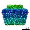 2233MC 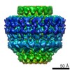 2232C  2ypwC  3zbjC C: citing same article ( M: map data used to model this data |
|---|---|
| Similar structure data |
-Links
 PDBj
PDBj


- Assembly
| Deposited unit | 
|
|---|---|
| 1 |
|
-Components
| #1: Protein | TNF receptor associated factor Mass: 22962.553 Da / Num. of mol.: 14 / Fragment: C-TERMINAL DOMAIN, RESIDUES 171-386 Source method: isolated from a genetically manipulated source Source: (gene. exp.) ESCHERICHIA COLI (E. coli) / Strain: BL21List of strains of Escherichia coli / Plasmid: PASK-IBA3C / Production host: ESCHERICHIA COLI (E. coli) / Strain (production host): BL21 / References: UniProt: Q46705#2: Protein | Mass: 14680.648 Da / Num. of mol.: 14 / Fragment: C-TERMINAL DOMAIN, RESIDUES 161-290 Source method: isolated from a genetically manipulated source Source: (gene. exp.) ESCHERICHIA COLI (E. coli) / Strain: BL21List of strains of Escherichia coli / Plasmid: PASK-IBA3C / Production host: ESCHERICHIA COLI (E. coli) / Strain (production host): BL21 / References: UniProt: Q46704#3: Protein/peptide | Mass: 5135.873 Da / Num. of mol.: 14 Source method: isolated from a genetically manipulated source Source: (gene. exp.) ESCHERICHIA COLI (E. coli) / Strain: BL21List of strains of Escherichia coli / Plasmid: PASK-IBA3C / Production host: ESCHERICHIA COLI (E. coli) / Strain (production host): BL21 / References: UniProt: Q46702 |
|---|
-Experimental details
-Experiment
| Experiment | Method: ELECTRON MICROSCOPY |
|---|---|
| EM experiment | Aggregation state: PARTICLE / 3D reconstruction method: single particle reconstruction |
- Sample preparation
| Component | Name: TRAN,TRAO AND TRAF COMPLEX ENCODED BY PKM101 DIGESTED WITH 0.02 MG ML- 1 OF ELASTASE FOR 40 MIN AT ROOM TEMPERATURE Type: COMPLEX |
|---|---|
| Buffer solution | Name: 50 MM TRIS-HCL, 200 MM NACL, 10 MM LDAO / pH: 8 / Details: 50 MM TRIS-HCL, 200 MM NACL, 10 MM LDAO |
| Specimen | Conc.: 0.01 mg/ml / Embedding applied: NO / Shadowing applied: NO / Staining applied: NO / Vitrification applied: YES |
| Specimen support | Details: CARBON |
| Vitrification | Instrument: FEI VITROBOT MARK III / Cryogen name: ETHANE Details: VITRIFICATION 1 -- CRYOGEN- ETHANE, HUMIDITY- 100, TEMPERATURE- 92, INSTRUMENT- FEI VITROBOT MARK III, METHOD- BLOT 4 SECONDS BEFORE PLUNGING, |
- Electron microscopy imaging
| Microscopy | Model: FEI TECNAI 12 / Date: Jan 11, 2012 / Details: 4000X4000 CCD |
|---|---|
| Electron gun | Electron source: FIELD EMISSION GUN / Accelerating voltage: 200 kV / Illumination mode: FLOOD BEAM |
| Electron lens | Mode: BRIGHT FIELDBright-field microscopy / Nominal magnification: 66000 X / Calibrated magnification: 68100 X / Nominal defocus max: 3500 nm / Nominal defocus min: 1500 nm / Cs: 2.1 mm |
| Specimen holder | Temperature: 95 K |
| Image recording | Electron dose: 20 e/Å2 / Film or detector model: GENERIC GATAN |
| Image scans | Num. digital images: 262 |
| Radiation wavelength | Relative weight: 1 |
- Processing
| EM software |
| ||||||||||||||||||
|---|---|---|---|---|---|---|---|---|---|---|---|---|---|---|---|---|---|---|---|
| CTF correction | Details: PHASE FLIPPING, EACH CCD IMAGE | ||||||||||||||||||
| Symmetry | Point symmetry: C14 (14 fold cyclic) | ||||||||||||||||||
| 3D reconstruction | Method: COMMON LINES / Resolution: 8.5 Å / Num. of particles: 5430 / Nominal pixel size: 2.2 Å / Actual pixel size: 2.08 Å Details: SUBMISSION BASED ON EXPERIMENTAL DATA FROM EMDB EMD -2233. (DEPOSITION ID: 11228). Symmetry type: POINT | ||||||||||||||||||
| Atomic model building | Protocol: FLEXIBLE FIT / Space: REAL / Target criteria: Cross-correlation coefficient Details: METHOD--RIGID BODY FOLLOWED BY FLEXIBLE FITTING REFINEMENT PROTOCOL--X-RAY | ||||||||||||||||||
| Atomic model building | PDB-ID: 3JQO | ||||||||||||||||||
| Refinement | Highest resolution: 8.5 Å | ||||||||||||||||||
| Refinement step | Cycle: LAST / Highest resolution: 8.5 Å
|

