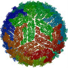
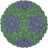
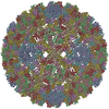
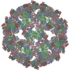
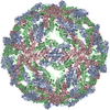
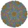
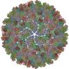
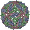
Journal: Proc Natl Acad Sci U S A / Year: 2013 Title: Dengue structure differs at the temperatures of its human and mosquito hosts. Authors: Xinzheng Zhang / Ju Sheng / Pavel Plevka / Richard J Kuhn / Michael S Diamond / Michael G Rossmann / Abstract: We report on a conformational transition of dengue virus when changing the temperature from that present in its mosquito vectors to that of its human host. Using cryoelectron microscopy, we show that ...We report on a conformational transition of dengue virus when changing the temperature from that present in its mosquito vectors to that of its human host. Using cryoelectron microscopy, we show that although the virus has a smooth surface, a diameter of ∼500 Å, and little exposed membrane at room temperature, the virions have a bumpy appearance with a diameter of ∼550 Å and some exposed membrane at 37 °C. The bumpy structure at 37 °C was found to be similar to the previously predicted structure of an intermediate between the smooth mature and fusogenic forms. As humans have a body temperature of 37 °C, the bumpy form of the virus would be the form present in humans. Thus, optimal dengue virus vaccines should induce antibodies that preferentially recognize epitopes exposed on the bumpy form of the virus.
In the structure databanks used in Yorodumi, some data are registered as the other names, "COVID-19 virus" and "2019-nCoV". Here are the details of the virus and the list of structure data.
Jan 31, 2019. EMDB accession codes are about to change! (news from PDBe EMDB page)
EMDB accession codes are about to change! (news from PDBe EMDB page)
The allocation of 4 digits for EMDB accession codes will soon come to an end. Whilst these codes will remain in use, new EMDB accession codes will include an additional digit and will expand incrementally as the available range of codes is exhausted. The current 4-digit format prefixed with “EMD-” (i.e. EMD-XXXX) will advance to a 5-digit format (i.e. EMD-XXXXX), and so on. It is currently estimated that the 4-digit codes will be depleted around Spring 2019, at which point the 5-digit format will come into force.
The EM Navigator/Yorodumi systems omit the EMD- prefix.
Related info.:Q: What is EMD? / ID/Accession-code notation in Yorodumi/EM Navigator
Yorodumi is a browser for structure data from EMDB, PDB, SASBDB, etc.
This page is also the successor to EM Navigator detail page, and also detail information page/front-end page for Omokage search.
The word "yorodu" (or yorozu) is an old Japanese word meaning "ten thousand". "mi" (miru) is to see.
Related info.:EMDB / PDB / SASBDB / Comparison of 3 databanks / Yorodumi Search / Aug 31, 2016. New EM Navigator & Yorodumi / Yorodumi Papers / Jmol/JSmol / Function and homology information / Changes in new EM Navigator and Yorodumi
 Movie
Movie Controller


 Open data
 Basic information
 Components
 Viral envelope
  Keywords
 Function and homology information
 Authors
 Citation
 Structure visualization
 Downloads & links
 Other downloads
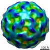
 PDBj
PDBj

 Assembly
 Sample preparation
 Electron microscopy imaging
 Processing