[English] 日本語
Yorodumi
- PDB-1n03: Model for Active RecA Filament -

+
Open data


ID or keywords:

Loading...

-
Basic information

Entry
Database: PDB / ID: 1n03
TitleModel for Active RecA Filament
ComponentsRecA protein
KeywordsDNA BINDING PROTEIN / helical polymer
Function / homology
Function and homology information


DNA polymerase V complex / homologous recombination / recombinational repair / SOS response / ATP-dependent DNA damage sensor activity / response to ionizing radiation / translesion synthesis / ATP-dependent activity, acting on DNA / cell motility / single-stranded DNA binding ...DNA polymerase V complex / homologous recombination / recombinational repair / SOS response / ATP-dependent DNA damage sensor activity / response to ionizing radiation / translesion synthesis / ATP-dependent activity, acting on DNA / cell motility / single-stranded DNA binding / DNA-binding transcription factor binding / DNA recombination / damaged DNA binding / DNA damage response / ATP hydrolysis activity / ATP binding / cytoplasm
Similarity search - Function
: / : / RecA C-terminal domain / DNA recombination/repair protein RecA, conserved site / DNA recombination and repair protein RecA, C-terminal / recA signature. / DNA recombination and repair protein RecA / recA bacterial DNA recombination protein / DNA recombination and repair protein RecA, monomer-monomer interface / RecA family profile 2. ...: / : / RecA C-terminal domain / DNA recombination/repair protein RecA, conserved site / DNA recombination and repair protein RecA, C-terminal / recA signature. / DNA recombination and repair protein RecA / recA bacterial DNA recombination protein / DNA recombination and repair protein RecA, monomer-monomer interface / RecA family profile 2. / DNA recombination and repair protein RecA-like, ATP-binding domain / RecA family profile 1. / ATPases associated with a variety of cellular activities / AAA+ ATPase domain / P-loop containing nucleoside triphosphate hydrolase
Similarity search - Domain/homology
ADENOSINE-5'-DIPHOSPHATE / Protein RecA
Similarity search - Component
Biological speciesEscherichia coli (E. coli)
MethodELECTRON MICROSCOPY / helical reconstruction / negative staining / Resolution: 20 Å
AuthorsVanLoock, M.S. / Yu, X. / Yang, S. / Lai, A.L. / Low, C. / Campbell, M.J. / Egelman, E.H.
Citation
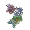
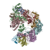
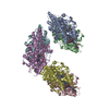
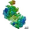
Journal: Structure / Year: 2003
Title: ATP-mediated conformational changes in the RecA filament.
Authors: Margaret S VanLoock / Xiong Yu / Shixin Yang / Alex L Lai / Claudia Low / Michael J Campbell / Edward H Egelman /
Abstract: The crystal structure of the E. coli RecA protein was solved more than 10 years ago, but it has provided limited insight into the mechanism of homologous genetic recombination. Using electron ...The crystal structure of the E. coli RecA protein was solved more than 10 years ago, but it has provided limited insight into the mechanism of homologous genetic recombination. Using electron microscopy, we have reconstructed five different states of RecA-DNA filaments. The C-terminal lobe of the RecA protein is modulated by the state of the distantly bound nucleotide, and this allosteric coupling can explain how mutations and truncations of this C-terminal lobe enhance RecA's activity. A model generated from these reconstructions shows that the nucleotide binding core is substantially rotated from its position in the RecA crystal filament, resulting in ATP binding between subunits. This simple rotation can explain the large cooperativity in ATP hydrolysis observed for RecA-DNA filaments.
History
DepositionOct 10, 2002Deposition site: RCSB / Processing site: RCSB
Revision 1.0Feb 25, 2003Provider: repository / Type: Initial release
Revision 1.1Apr 28, 2008Group: Version format compliance
Revision 1.2Jul 13, 2011Group: Version format compliance
Revision 1.3Jul 18, 2018Group: Author supporting evidence / Data collection / Category: em_single_particle_entity / em_software / Item: _em_software.image_processing_id
Revision 1.4Nov 6, 2019Group: Advisory / Data collection / Other / Category: atom_sites / cell / pdbx_validate_symm_contact
Item: _atom_sites.fract_transf_matrix[1][1] / _atom_sites.fract_transf_matrix[2][2] ..._atom_sites.fract_transf_matrix[1][1] / _atom_sites.fract_transf_matrix[2][2] / _atom_sites.fract_transf_matrix[3][3] / _cell.Z_PDB / _cell.angle_alpha / _cell.angle_beta / _cell.angle_gamma / _cell.length_a / _cell.length_b / _cell.length_c
Revision 1.5Feb 14, 2024Group: Data collection / Database references ...Data collection / Database references / Derived calculations / Refinement description
Category: chem_comp_atom / chem_comp_bond ...chem_comp_atom / chem_comp_bond / database_2 / em_3d_fitting_list / pdbx_initial_refinement_model / struct_site
Item: _database_2.pdbx_DOI / _database_2.pdbx_database_accession ..._database_2.pdbx_DOI / _database_2.pdbx_database_accession / _em_3d_fitting_list.accession_code / _em_3d_fitting_list.initial_refinement_model_id / _em_3d_fitting_list.source_name / _em_3d_fitting_list.type / _struct_site.pdbx_auth_asym_id / _struct_site.pdbx_auth_comp_id / _struct_site.pdbx_auth_seq_id

-
Structure visualization

Movie
  • Deposited structure unit
  • Imaged by Jmol
  • Download
Movie viewer
Structure viewerMolecule:
MolmilJmol/JSmol

Downloads & links

-
Assembly

Deposited unit
A: RecA protein
B: RecA protein
C: RecA protein
D: RecA protein
E: RecA protein
F: RecA protein
G: RecA protein
hetero molecules


Theoretical massNumber of molelcules
Total (without water)268,18614
Polymers265,1967
Non-polymers2,9907
Water0
1


  • Idetical with deposited unit
  • defined by author
TypeNameSymmetry operationNumber
identity operation1_555x,y,z1

-
Components

#1: Protein
RecA protein / / Recombinase A / Coordinate model: Cα atoms only


Mass: 37885.086 Da / Num. of mol.: 7 / Source method: isolated from a natural source / Source: (natural) Escherichia coli (E. coli) / References: UniProt: P0A7G6
#2: Chemical
ChemComp-ADP / ADENOSINE-5'-DIPHOSPHATE / Adenosine diphosphate


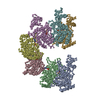
Mass: 427.201 Da / Num. of mol.: 7 / Source method: obtained synthetically / Formula: C10H15N5O10P2 / Comment: ADP, energy-carrying molecule*YM

-
Experimental details

-
Experiment

ExperimentMethod: ELECTRON MICROSCOPY
EM experimentAggregation state: FILAMENT / 3D reconstruction method: helical reconstruction

-
Sample preparation

ComponentName: RecA Filament / Type: COMPLEX
Details: The active form of the RecA protein is the helical polymer formed on DNA in the presence of ATP
SpecimenEmbedding applied: NO / Shadowing applied: NO / Staining applied: YES / Vitrification applied: NO
EM stainingType: NEGATIVE / Material: Uranyl Acetate
Crystal grow
*PLUS
Method: electron microscopy / Details: Electron Microscopy

-
Electron microscopy imaging

MicroscopyModel: FEI TECNAI 12
Electron gunElectron source: OTHER / Accelerating voltage: 80 kV / Illumination mode: FLOOD BEAM
Electron lensMode: BRIGHT FIELDBright-field microscopy / Nominal magnification: 30000 X
Image recordingFilm or detector model: GENERIC FILM
Image scansScanner model: OTHER

-
Processing

EM software
IDNameCategory
1Omodel fitting
2IHRSR3D reconstruction
3SPIDER3D reconstruction
3D reconstructionMethod: Iterative Helical Real Space Reconstruction method (Egelman, Ultramicroscopy 85, 225-234, 2000) was used.
Resolution: 20 Å / Num. of particles: 60000 / Nominal pixel size: 3.9 Å / Actual pixel size: 3.9 Å / Magnification calibration: TMV PARTICLES / Symmetry type: HELICAL
Atomic model buildingProtocol: RIGID BODY FIT / Space: REAL / Target criteria: best visual fit using the program O / Details: REFINEMENT PROTOCOL--rigid body
Atomic model buildingPDB-ID: 1REA
Accession code: 1REA / Source name: PDB / Type: experimental model
Refinement stepCycle: LAST
ProteinNucleic acidLigandSolventTotal
Num. atoms2121 0 189 0 2310

+
About Yorodumi

-
News

-
Feb 9, 2022. New format data for meta-information of EMDB entries

New format data for meta-information of EMDB entries

  • Version 3 of the EMDB header file is now the official format.
  • The previous official version 1.9 will be removed from the archive.

Related info.:EMDB header

External links:wwPDB to switch to version 3 of the EMDB data model

-
Aug 12, 2020. Covid-19 info

Covid-19 info

URL: https://pdbj.org/emnavi/covid19.php

New page: Covid-19 featured information page in EM Navigator.

Related info.:Covid-19 info / Mar 5, 2020. Novel coronavirus structure data

+
Mar 5, 2020. Novel coronavirus structure data

Novel coronavirus structure data

Related info.:Yorodumi Speices / Aug 12, 2020. Covid-19 info

External links:COVID-19 featured content - PDBj / Molecule of the Month (242):Coronavirus Proteases

+
Jan 31, 2019. EMDB accession codes are about to change! (news from PDBe EMDB page)

EMDB accession codes are about to change! (news from PDBe EMDB page)

  • The allocation of 4 digits for EMDB accession codes will soon come to an end. Whilst these codes will remain in use, new EMDB accession codes will include an additional digit and will expand incrementally as the available range of codes is exhausted. The current 4-digit format prefixed with “EMD-” (i.e. EMD-XXXX) will advance to a 5-digit format (i.e. EMD-XXXXX), and so on. It is currently estimated that the 4-digit codes will be depleted around Spring 2019, at which point the 5-digit format will come into force.
  • The EM Navigator/Yorodumi systems omit the EMD- prefix.

Related info.:Q: What is EMD? / ID/Accession-code notation in Yorodumi/EM Navigator

External links:EMDB Accession Codes are Changing Soon! / Contact to PDBj

+
Jul 12, 2017. Major update of PDB

Major update of PDB

  • wwPDB released updated PDB data conforming to the new PDBx/mmCIF dictionary.
  • This is a major update changing the version number from 4 to 5, and with Remediation, in which all the entries are updated.
  • In this update, many items about electron microscopy experimental information are reorganized (e.g. em_software).
  • Now, EM Navigator and Yorodumi are based on the updated data.

External links:wwPDB Remediation / Enriched Model Files Conforming to OneDep Data Standards Now Available in the PDB FTP Archive

-
Yorodumi

Thousand views of thousand structures

  • Yorodumi is a browser for structure data from EMDB, PDB, SASBDB, etc.
  • This page is also the successor to EM Navigator detail page, and also detail information page/front-end page for Omokage search.
  • The word "yorodu" (or yorozu) is an old Japanese word meaning "ten thousand". "mi" (miru) is to see.

Related info.:EMDB / PDB / SASBDB / Comparison of 3 databanks / Yorodumi Search / Aug 31, 2016. New EM Navigator & Yorodumi / Yorodumi Papers / Jmol/JSmol / Function and homology information / Changes in new EM Navigator and Yorodumi

Read more