 Movie
Movie Controller

[English] 日本語
 Yorodumi
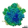
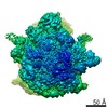
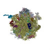
- EMDB-8369: Methicillin Resistant, Linezolid resistant Staphylococcus aureus ... -
+ Open data

- Basic information
| Entry | Database: EMDB / ID: EMD-8369 | |||||||||
|---|---|---|---|---|---|---|---|---|---|---|
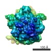
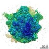
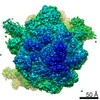
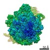
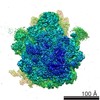
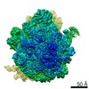
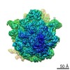
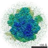
| Title | Methicillin Resistant, Linezolid resistant Staphylococcus aureus 70S ribosome (delta S145 uL3) | |||||||||
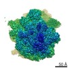
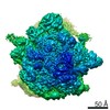
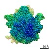
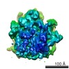
 Map data Map data | ||||||||||
 Sample Sample |
| |||||||||
| Function / homology |  Function and homology information Function and homology informationlarge ribosomal subunit / small ribosomal subunit /  transferase activity / tRNA binding / rRNA binding / ribosome / structural constituent of ribosome / ribonucleoprotein complex / translation / cytoplasm transferase activity / tRNA binding / rRNA binding / ribosome / structural constituent of ribosome / ribonucleoprotein complex / translation / cytoplasmSimilarity search - Function | |||||||||
| Biological species |   Staphylococcus aureus (bacteria) Staphylococcus aureus (bacteria) | |||||||||
| Method | single particle reconstruction / cryo EM / Resolution: 3.6 Å | |||||||||
 Authors Authors | Belousoff MJ / Lithgow T / Eyal Z / Yonath A / Radjainia M | |||||||||
| Funding support |  Australia, 1 items Australia, 1 items
| |||||||||
 Citation Citation | Journal: mBio / Year: 2017 Title: Structural Basis for Linezolid Binding Site Rearrangement in the Ribosome. Authors: Matthew J Belousoff / Zohar Eyal / Mazdak Radjainia / Tofayel Ahmed / Rebecca S Bamert / Donna Matzov / Anat Bashan / Ella Zimmerman / Satabdi Mishra / David Cameron / Hans Elmlund / Anton Y ...Authors: Matthew J Belousoff / Zohar Eyal / Mazdak Radjainia / Tofayel Ahmed / Rebecca S Bamert / Donna Matzov / Anat Bashan / Ella Zimmerman / Satabdi Mishra / David Cameron / Hans Elmlund / Anton Y Peleg / Shashi Bhushan / Trevor Lithgow / Ada Yonath /   Abstract: An unorthodox, surprising mechanism of resistance to the antibiotic linezolid was revealed by cryo-electron microscopy (cryo-EM) in the 70S ribosomes from a clinical isolate of This high-resolution ...An unorthodox, surprising mechanism of resistance to the antibiotic linezolid was revealed by cryo-electron microscopy (cryo-EM) in the 70S ribosomes from a clinical isolate of This high-resolution structural information demonstrated that a single amino acid deletion in ribosomal protein uL3 confers linezolid resistance despite being located 24 Å away from the linezolid binding pocket in the peptidyl-transferase center. The mutation induces a cascade of allosteric structural rearrangements of the rRNA that ultimately results in the alteration of the antibiotic binding site. The growing burden on human health caused by various antibiotic resistance mutations now includes prevalent resistance to last-line antimicrobial drugs such as linezolid and daptomycin. Structure-informed drug modification represents a frontier with respect to designing advanced clinical therapies, but success in this strategy requires rapid, facile means to shed light on the structural basis for drug resistance (D. Brown, Nat Rev Drug Discov 14:821-832, 2015, https://doi.org/10.1038/nrd4675). Here, detailed structural information demonstrates that a common mechanism is at play in linezolid resistance and provides a step toward the redesign of oxazolidinone antibiotics, a strategy that could thwart known mechanisms of linezolid resistance. | |||||||||
| History |
|
- Structure visualization
| Movie |
Movie viewer |
|---|---|
| Structure viewer | EM map: SurfViewMolmilJmol/JSmol |
| Supplemental images |

- Downloads & links
-EMDB archive
| Map data | emd_8369.map.gz | 17.1 MB | EMDB map data format | |
|---|---|---|---|---|
| Header (meta data) | emd-8369-v30.xmlemd-8369.xml | 49.9 KB 49.9 KB | Display Display | EMDB header |
| Images | 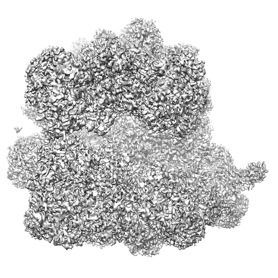 emd_8369.png emd_8369.png | 108 KB | ||
| Archive directory |  http://ftp.pdbj.org/pub/emdb/structures/EMD-8369ftp://ftp.pdbj.org/pub/emdb/structures/EMD-8369 http://ftp.pdbj.org/pub/emdb/structures/EMD-8369ftp://ftp.pdbj.org/pub/emdb/structures/EMD-8369 | HTTPS FTP |
-Related structure data
| Related structure data | 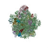 5t7vMC 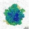 8402C 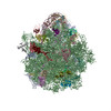 5tcuC M: atomic model generated by this map C: citing same article ( |
|---|---|
| Similar structure data |
-Links
| EMDB pages | EMDB (EBI/PDBe) / EMDataResource |
|---|---|
| Related items in Molecule of the Month |


-Map
| File | Download / File: emd_8369.map.gz / Format: CCP4 / Size: 125 MB / Type: IMAGE STORED AS FLOATING POINT NUMBER (4 BYTES) | ||||||||||||||||||||||||||||||||||||||||||||||||||||||||||||
|---|---|---|---|---|---|---|---|---|---|---|---|---|---|---|---|---|---|---|---|---|---|---|---|---|---|---|---|---|---|---|---|---|---|---|---|---|---|---|---|---|---|---|---|---|---|---|---|---|---|---|---|---|---|---|---|---|---|---|---|---|---|
| Voxel size | X=Y=Z: 1.1 Å | ||||||||||||||||||||||||||||||||||||||||||||||||||||||||||||
| Density |
| ||||||||||||||||||||||||||||||||||||||||||||||||||||||||||||
| Symmetry | Space group: 1 | ||||||||||||||||||||||||||||||||||||||||||||||||||||||||||||
| Details | EMDB XML:
CCP4 map header:
| ||||||||||||||||||||||||||||||||||||||||||||||||||||||||||||
-Supplemental data
- Sample components
+Entire : Clinical isolate Linezolid resistant MRSA 70S ribosome
+Supramolecule #1: Clinical isolate Linezolid resistant MRSA 70S ribosome
+Macromolecule #1: 16S ribosomal RNA
+Macromolecule #14: 23S ribosomal RNA
+Macromolecule #15: 5S ribosomal RNA
+Macromolecule #42: E-site tRNA
+Macromolecule #2: 30S ribosomal protein S10
+Macromolecule #3: 30S ribosomal protein S11
+Macromolecule #4: 30S ribosomal protein S12
+Macromolecule #5: 30S ribosomal protein S15
+Macromolecule #6: 30S ribosomal protein S16
+Macromolecule #7: 30S ribosomal protein S17
+Macromolecule #8: 30S ribosomal protein S18
+Macromolecule #9: 30S ribosomal protein S20
+Macromolecule #10: 30S ribosomal protein S4
+Macromolecule #11: 30S ribosomal protein S5
+Macromolecule #12: 30S ribosomal protein S6
+Macromolecule #13: 30S ribosomal protein S8
+Macromolecule #16: 50S ribosomal protein L19
+Macromolecule #17: 50S ribosomal protein L2
+Macromolecule #18: 50S ribosomal protein L20
+Macromolecule #19: 50S ribosomal protein L21
+Macromolecule #20: 50S ribosomal protein L22
+Macromolecule #21: 50S ribosomal protein L23
+Macromolecule #22: 50S ribosomal protein L24
+Macromolecule #23: 50S ribosomal protein L25
+Macromolecule #24: 50S ribosomal protein L27
+Macromolecule #25: 50S ribosomal protein L28
+Macromolecule #26: 50S ribosomal protein L29
+Macromolecule #27: 50S ribosomal protein L3
+Macromolecule #28: 50S ribosomal protein L30
+Macromolecule #29: 50S ribosomal protein L32
+Macromolecule #30: 50S ribosomal protein L33
+Macromolecule #31: 50S ribosomal protein L34
+Macromolecule #32: 50S ribosomal protein L35
+Macromolecule #33: 50S ribosomal protein L36
+Macromolecule #34: 50S ribosomal protein L4
+Macromolecule #35: 50S ribosomal protein L6
+Macromolecule #36: 50S ribosomal protein L13
+Macromolecule #37: 50S ribosomal protein L14
+Macromolecule #38: 50S ribosomal protein L15
+Macromolecule #39: 50S ribosomal protein L16
+Macromolecule #40: 50S ribosomal protein L17
+Macromolecule #41: 50S ribosomal protein L18
+Macromolecule #43: MAGNESIUM ION
-Experimental details
-Structure determination
| Method | cryo EM |
|---|---|
 Processing Processing | single particle reconstruction |
| Aggregation state | particle |
-Sample preparation
| Concentration | 0.3 mg/mL |
|---|---|
| Buffer | pH: 7.5 |
| Grid | Model: Quantifoil R1.2/1.3 / Material: COPPER / Mesh: 300 / Support film - Material: CARBON / Support film - topology: HOLEY ARRAY |
| Vitrification | Cryogen name: ETHANE / Chamber humidity: 100 % / Chamber temperature: 277 K / Instrument: FEI VITROBOT MARK IV |
- Electron microscopy
| Microscope | FEI TITAN KRIOS |
|---|---|
| Electron beam | Acceleration voltage: 300 kV / Electron source: FIELD EMISSION GUN |
| Electron optics | Illumination mode: SPOT SCAN / Imaging mode: BRIGHT FIELDBright-field microscopy |
| Image recording | Film or detector model: FEI FALCON II (4k x 4k) / Average electron dose: 45.0 e/Å2 |
| Experimental equipment |  Model: Titan Krios / Image courtesy: FEI Company |
-Image processing
| Startup model | Type of model: PDB ENTRY PDB model - PDB ID: |
|---|---|
| Initial angle assignment | Type: PROJECTION MATCHING |
| Final angle assignment | Type: ANGULAR RECONSTITUTION |
| Final reconstruction | Resolution.type: BY AUTHOR / Resolution: 3.6 Å / Resolution method: FSC 0.143 CUT-OFF / Number images used: 80500 |
-Atomic model buiding 1
| Refinement | Space: REAL / Protocol: FLEXIBLE FIT |
|---|---|
| Output model | PDB-5t7v: |
