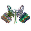
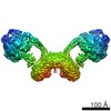
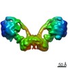
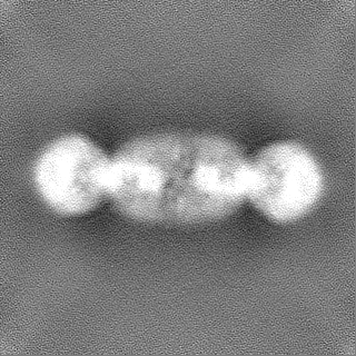
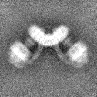
- EMDB-7067: Cryo-EM structure of dimeric F1FO yeast mitochondrial ATP synthas... -
+
Open data
ID or keywords:
Loading...
-
Basic information
Entry
Database: EMDB / ID: EMD-7067
Title
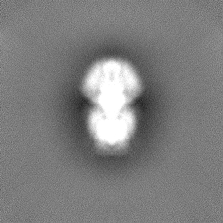
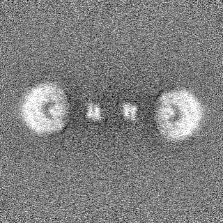
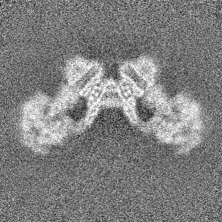
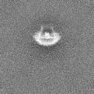
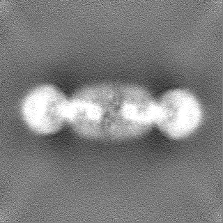
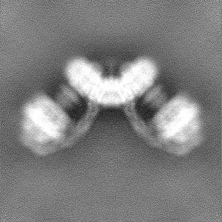
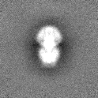
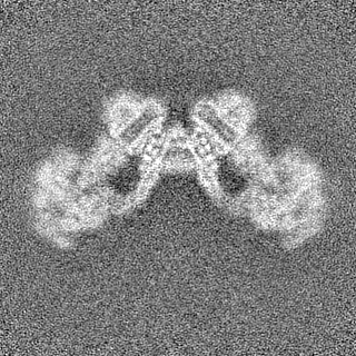
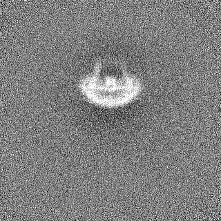
Cryo-EM structure of dimeric F1FO yeast mitochondrial ATP synthase with C2 symmetry
Map data
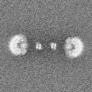
Sharpened map of the dimeric FO region of yeast mitochondrial ATP synthase
Sample
Complex: Yeast mitochondrial F1Fo ATP synthase dimer
Function / homology
Function and homology information
mitochondrial proton-transporting ATP synthase, central stalk / mitochondrial proton-transporting ATP synthase complex assembly / mitochondrial proton-transporting ATP synthase, catalytic core / mitochondrial proton-transporting ATP synthase, stator stalk / mitochondrial proton-transporting ATP synthase complex, coupling factor F(o) / mitochondrial proton-transporting ATP synthase complex / mitochondrial proton-transporting ATP synthase complex, catalytic sector F(1) / mitochondrial nucleoid / proton motive force-driven mitochondrial ATP synthesis / proton motive force-driven ATP synthesis ...mitochondrial proton-transporting ATP synthase, central stalk / mitochondrial proton-transporting ATP synthase complex assembly / mitochondrial proton-transporting ATP synthase, catalytic core / mitochondrial proton-transporting ATP synthase, stator stalk / mitochondrial proton-transporting ATP synthase complex, coupling factor F(o) / mitochondrial proton-transporting ATP synthase complex / mitochondrial proton-transporting ATP synthase complex, catalytic sector F(1) / mitochondrial nucleoid / proton motive force-driven mitochondrial ATP synthesis / proton motive force-driven ATP synthesis / proton transmembrane transporter activity / proton-transporting ATP synthase complex, catalytic core F(1) / H+-transporting two-sector ATPase / proton-transporting ATPase activity, rotational mechanism / proton transmembrane transport / proton-transporting ATP synthase activity, rotational mechanism / ADP binding / mitochondrial intermembrane space / protein-containing complex assembly / mitochondrial inner membrane / lipid binding / ATP hydrolysis activity / mitochondrion / ATP binding / identical protein binding / cytosol Similarity search - Function
ATP synthase subunit K / ATP synthase subunit K / ATP synthase, F0 complex, subunit J / ATP synthase protein 8, fungal type / ATP synthase, F0 complex, subunit F, mitochondria, fungi / ATP synthase j chain / Fungal ATP synthase protein 8 (A6L) / Mitochondrial F1-F0 ATP synthase subunit F of fungi / : / Fungal epsilon subunit of F1F0-ATP synthase C-terminal domain ...ATP synthase subunit K / ATP synthase subunit K / ATP synthase, F0 complex, subunit J / ATP synthase protein 8, fungal type / ATP synthase, F0 complex, subunit F, mitochondria, fungi / ATP synthase j chain / Fungal ATP synthase protein 8 (A6L) / Mitochondrial F1-F0 ATP synthase subunit F of fungi / : / Fungal epsilon subunit of F1F0-ATP synthase C-terminal domain / ATP synthase, F0 complex, subunit B/MI25 / ATP synthase, F0 complex, subunit B / Mitochondrial ATP synthase B chain precursor (ATP-synt_B) / ATP synthase, F0 complex, subunit D, mitochondrial / ATP synthase D chain, mitochondrial (ATP5H) / ATP synthase, F0 complex, subunit D superfamily, mitochondrial / ATP synthase, F0 complex, subunit A, bacterial/mitochondria / ATP synthase, F1 complex, epsilon subunit, mitochondrial / ATP synthase, F1 complex, epsilon subunit superfamily, mitochondrial / Mitochondrial ATP synthase epsilon chain / ATPase, OSCP/delta subunit, conserved site / ATP synthase delta (OSCP) subunit signature. / F1F0 ATP synthase OSCP/delta subunit, N-terminal domain superfamily / ATP synthase, F0 complex, subunit A / ATP synthase, F0 complex, subunit A, active site / ATP synthase, F0 complex, subunit A superfamily / ATP synthase A chain / ATP synthase a subunit signature. / ATPase, OSCP/delta subunit / ATP synthase delta (OSCP) subunit / ATP synthase, F1 complex, delta/epsilon subunit / ATP synthase, F1 complex, delta/epsilon subunit, N-terminal / F0F1 ATP synthase delta/epsilon subunit, N-terminal / ATP synthase, Delta/Epsilon chain, beta-sandwich domain / ATP synthase, F0 complex, subunit C / F1F0 ATP synthase subunit C superfamily / ATP synthase, F0 complex, subunit C, DCCD-binding site / ATP synthase c subunit signature. / ATP synthase, F1 complex, gamma subunit conserved site / ATP synthase gamma subunit signature. / ATP synthase, F1 complex, beta subunit / ATP synthase, alpha subunit, C-terminal domain superfamily / ATP synthase, F1 complex, gamma subunit / ATP synthase, F1 complex, gamma subunit superfamily / ATP synthase / ATP synthase, alpha subunit, C-terminal / ATP synthase, F1 complex, alpha subunit / ATP synthase, F1 complex, alpha subunit nucleotide-binding domain / ATP synthase alpha/beta chain, C terminal domain / V-ATPase proteolipid subunit C-like domain / F/V-ATP synthase subunit C superfamily / ATP synthase subunit C / ATPase, F1/V1 complex, beta/alpha subunit, C-terminal / ATP synthase subunit alpha, N-terminal domain-like superfamily / ATPase, F1/V1/A1 complex, alpha/beta subunit, N-terminal domain superfamily / ATPase, F1/V1/A1 complex, alpha/beta subunit, N-terminal domain / ATP synthase alpha/beta family, beta-barrel domain / ATPase, alpha/beta subunit, nucleotide-binding domain, active site / ATP synthase alpha and beta subunits signature. / ATPase, F1/V1/A1 complex, alpha/beta subunit, nucleotide-binding domain / ATP synthase alpha/beta family, nucleotide-binding domain / ATPases associated with a variety of cellular activities / AAA+ ATPase domain / P-loop containing nucleoside triphosphate hydrolase Similarity search - Domain/homology
ATP synthase catalytic sector F1 epsilon subunit / ATP synthase subunit beta, mitochondrial / ATP synthase subunit a / ATP synthase protein 8 / ATP synthase subunit 4, mitochondrial / ATP synthase subunit alpha, mitochondrial / ATP synthase subunit 5, mitochondrial / ATP synthase subunit d, mitochondrial / ATP synthase subunit gamma, mitochondrial / ATP synthase subunit 9, mitochondrial ...ATP synthase catalytic sector F1 epsilon subunit / ATP synthase subunit beta, mitochondrial / ATP synthase subunit a / ATP synthase protein 8 / ATP synthase subunit 4, mitochondrial / ATP synthase subunit alpha, mitochondrial / ATP synthase subunit 5, mitochondrial / ATP synthase subunit d, mitochondrial / ATP synthase subunit gamma, mitochondrial / ATP synthase subunit 9, mitochondrial / ATP synthase subunit J, mitochondrial / ATP synthase subunit K, mitochondrial / ATP synthase subunit f, mitochondrial / ATP synthase subunit delta, mitochondrial Similarity search - Component
Biological species
Saccharomyces cerevisiae (brewer's yeast)
Method
single particle reconstruction / cryo EM / Resolution: 7.4 Å
NIH National Institute of General Medical Sciences
GM103310
United States
Citation
Journal: Science / Year: 2017 Title: Atomic model for the dimeric F region of mitochondrial ATP synthase. Authors: Hui Guo / Stephanie A Bueler / John L Rubinstein / Abstract: Mitochondrial adenosine triphosphate (ATP) synthase produces the majority of ATP in eukaryotic cells, and its dimerization is necessary to create the inner membrane folds, or cristae, characteristic ...Mitochondrial adenosine triphosphate (ATP) synthase produces the majority of ATP in eukaryotic cells, and its dimerization is necessary to create the inner membrane folds, or cristae, characteristic of mitochondria. Proton translocation through the membrane-embedded F region turns the rotor that drives ATP synthesis in the soluble F region. Although crystal structures of the F region have illustrated how this rotation leads to ATP synthesis, understanding how proton translocation produces the rotation has been impeded by the lack of an experimental atomic model for the F region. Using cryo-electron microscopy, we determined the structure of the dimeric F complex from at a resolution of 3.6 angstroms. The structure clarifies how the protons travel through the complex, how the complex dimerizes, and how the dimers bend the membrane to produce cristae.
History
Deposition
Oct 7, 2017
-
Header (metadata) release
Nov 8, 2017
-
Map release
Nov 8, 2017
-
Update
Nov 25, 2020
-
Current status
Nov 25, 2020
Processing site: RCSB / Status: Released
-
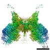
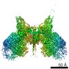
Structure visualization
Movie
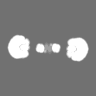
Surface view with section colored by density value
Film or detector model: GATAN K2 QUANTUM (4k x 4k) / Detector mode: COUNTING / Digitization - Frames/image: 1-30 / Number real images: 626 / Average exposure time: 15.0 sec. / Average electron dose: 36.0 e/Å2
Experimental equipment
Model: Tecnai F20 / Image courtesy: FEI Company
-
Image processing
Particle selection
Number selected: 143294
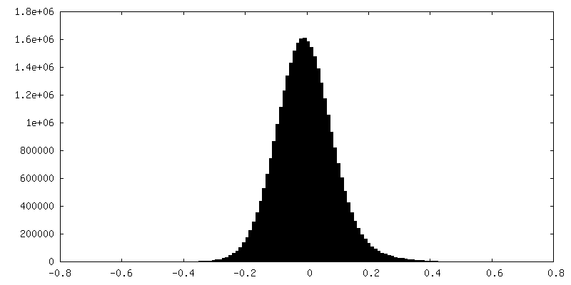
CTF correction
Software - Name: CTFFIND (ver. 4)
Startup model
Type of model: OTHER Details: Starting model generated by ab initial 3D reconstruction with cryoSPARC.
Initial angle assignment
Type: OTHER
Final angle assignment
Type: PROJECTION MATCHING
Final reconstruction
Number classes used: 1 / Applied symmetry - Point group: C2 (2 fold cyclic) / Resolution.type: BY AUTHOR / Resolution: 7.4 Å / Resolution method: FSC 0.143 CUT-OFF / Software - Name: cryoSPARC (ver. 0.4.1) / Number images used: 79942
-
Atomic model buiding 1
Refinement
Protocol: AB INITIO MODEL
Output model
PDB-6b8h: Mosaic model of yeast mitochondrial ATP synthase monomer
+
About Yorodumi
-
News
-
Feb 9, 2022. New format data for meta-information of EMDB entries
New format data for meta-information of EMDB entries
Version 3 of the EMDB header file is now the official format.
The previous official version 1.9 will be removed from the archive.
In the structure databanks used in Yorodumi, some data are registered as the other names, "COVID-19 virus" and "2019-nCoV". Here are the details of the virus and the list of structure data.
Jan 31, 2019. EMDB accession codes are about to change! (news from PDBe EMDB page)
EMDB accession codes are about to change! (news from PDBe EMDB page)
The allocation of 4 digits for EMDB accession codes will soon come to an end. Whilst these codes will remain in use, new EMDB accession codes will include an additional digit and will expand incrementally as the available range of codes is exhausted. The current 4-digit format prefixed with “EMD-” (i.e. EMD-XXXX) will advance to a 5-digit format (i.e. EMD-XXXXX), and so on. It is currently estimated that the 4-digit codes will be depleted around Spring 2019, at which point the 5-digit format will come into force.
The EM Navigator/Yorodumi systems omit the EMD- prefix.
Related info.:Q: What is EMD? / ID/Accession-code notation in Yorodumi/EM Navigator
Yorodumi is a browser for structure data from EMDB, PDB, SASBDB, etc.
This page is also the successor to EM Navigator detail page, and also detail information page/front-end page for Omokage search.
The word "yorodu" (or yorozu) is an old Japanese word meaning "ten thousand". "mi" (miru) is to see.
Related info.:EMDB / PDB / SASBDB / Comparison of 3 databanks / Yorodumi Search / Aug 31, 2016. New EM Navigator & Yorodumi / Yorodumi Papers / Jmol/JSmol / Function and homology information / Changes in new EM Navigator and Yorodumi
 Movie
Movie Controller

 Yorodumi
 Open data
 Basic information
 Map data
 Sample
 Function and homology information
 H+-transporting two-sector ATPase / proton-transporting ATPase activity, rotational mechanism / proton transmembrane transport / proton-transporting ATP synthase activity, rotational mechanism /
 Authors
 Canada,
  United States, 3 items
  Citation
 Structure visualization
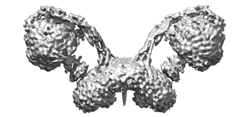
 Downloads & links
 emd_7067.png
 http://ftp.pdbj.org/pub/emdb/structures/EMD-7067
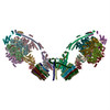


 Z
Z Y
Y X
X


 Sample components
 Processing
 Electron microscopy
