 Movie
Movie Controller

[English] 日本語
 Yorodumi
- EMDB-6675: CryoEM structure of type II secretion system secretin GspD in E.c... -
+ Open data

- Basic information
| Entry | Database: EMDB / ID: EMD-6675 | |||||||||
|---|---|---|---|---|---|---|---|---|---|---|
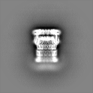
| Title | CryoEM structure of type II secretion system secretin GspD in E.coli K12 Type II secretion system Type II secretion system | |||||||||
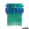
 Map data Map data | K12_GspD post-processing map 3.04 | |||||||||
 Sample Sample |
| |||||||||
 Keywords Keywords | Secretin family / C15 symmetry / T2SS / PROTEIN TRANSPORT | |||||||||
| Function / homology |  Function and homology informationprotein secretion by the type II secretion system / type II protein secretion system complex / protein secretion / cell outer membrane / membrane => GO:0016020 / identical protein binding Function and homology informationprotein secretion by the type II secretion system / type II protein secretion system complex / protein secretion / cell outer membrane / membrane => GO:0016020 / identical protein bindingSimilarity search - Function | |||||||||
| Biological species |  Escherichia coli K-12 (bacteria) Escherichia coli K-12 (bacteria) | |||||||||
| Method | single particle reconstruction / cryo EM / Resolution: 3.04 Å | |||||||||
 Authors Authors | Yan Z / Yin M | |||||||||
 Citation Citation | Journal: Nat Struct Mol Biol / Year: 2017 Title: Structural insights into the secretin translocation channel in the type II secretion system. Authors: Zhaofeng Yan / Meng Yin / Dandan Xu / Yongqun Zhu / Xueming Li /  Abstract: The secretin GspD of the type II secretion system (T2SS) forms a channel across the outer membrane in Gram-negative bacteria to transport substrates from the periplasm to the extracellular milieu. ...The secretin GspD of the type II secretion system (T2SS) forms a channel across the outer membrane in Gram-negative bacteria to transport substrates from the periplasm to the extracellular milieu. The lack of an atomic-resolution structure of the GspD channel hinders the investigation of substrate translocation mechanism of T2SS. Here we report cryo-EM structures of two GspD channels (∼1 MDa), from Escherichia coli K12 and Vibrio cholerae, at ∼3 Å resolution. The structures reveal a pentadecameric channel architecture, wherein three rings of GspD N domains form the periplasmic channel. The secretin domain constitutes a novel double β-barrel channel, with at least 60 β-strands in each barrel, and is stabilized by S domains. The outer membrane channel is sealed by β-strand-enriched gates. On the basis of the partially open state captured, we proposed a detailed gate-opening mechanism. Our structures provide a structural basis for understanding the secretin superfamily and the mechanism of substrate translocation in T2SS. | |||||||||
| History |
|
- Structure visualization
| Movie |
Movie viewer |
|---|---|
| Structure viewer | EM map: SurfViewMolmilJmol/JSmol |
| Supplemental images |

- Downloads & links
-EMDB archive
| Map data | emd_6675.map.gz | 192.7 MB | EMDB map data format | |
|---|---|---|---|---|
| Header (meta data) | emd-6675-v30.xmlemd-6675.xml | 12.3 KB 12.3 KB | Display Display | EMDB header |
| Images | 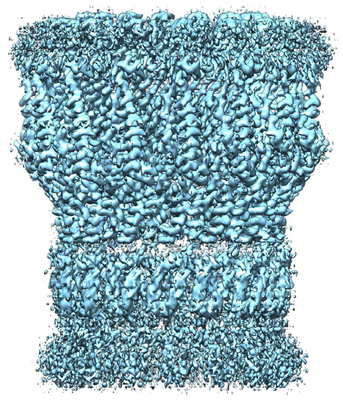 emd_6675.png emd_6675.png | 302.2 KB | ||
| Filedesc metadata | emd-6675.cif.gz | 5.3 KB | ||
| Others | emd_6675_additional.map.gz | 184.9 MB | ||
| Archive directory |  http://ftp.pdbj.org/pub/emdb/structures/EMD-6675ftp://ftp.pdbj.org/pub/emdb/structures/EMD-6675 http://ftp.pdbj.org/pub/emdb/structures/EMD-6675ftp://ftp.pdbj.org/pub/emdb/structures/EMD-6675 | HTTPS FTP |
-Related structure data
| Related structure data |  5wq7MC  6676C  6677C 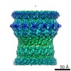 6678C  5wq8C 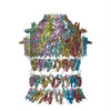 5wq9C M: atomic model generated by this map C: citing same article ( |
|---|---|
| Similar structure data |


-Links
| EMDB pages | EMDB (EBI/PDBe) / EMDataResource |
|---|---|
| Related items in Molecule of the Month |

-Map
| File | Download / File: emd_6675.map.gz / Format: CCP4 / Size: 209.3 MB / Type: IMAGE STORED AS FLOATING POINT NUMBER (4 BYTES) | ||||||||||||||||||||||||||||||||||||||||||||||||||||||||||||
|---|---|---|---|---|---|---|---|---|---|---|---|---|---|---|---|---|---|---|---|---|---|---|---|---|---|---|---|---|---|---|---|---|---|---|---|---|---|---|---|---|---|---|---|---|---|---|---|---|---|---|---|---|---|---|---|---|---|---|---|---|---|
| Annotation | K12_GspD post-processing map 3.04 | ||||||||||||||||||||||||||||||||||||||||||||||||||||||||||||
| Voxel size | X=Y=Z: 1.32 Å | ||||||||||||||||||||||||||||||||||||||||||||||||||||||||||||
| Density |
| ||||||||||||||||||||||||||||||||||||||||||||||||||||||||||||
| Symmetry | Space group: 1 | ||||||||||||||||||||||||||||||||||||||||||||||||||||||||||||
| Details | EMDB XML:
CCP4 map header:
| ||||||||||||||||||||||||||||||||||||||||||||||||||||||||||||
-Supplemental data
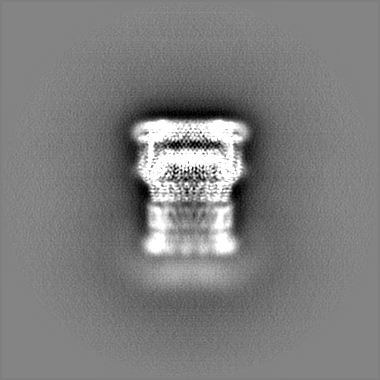
-Additional map: K12 GspD unpost-processing map
| File | emd_6675_additional.map | ||||||||||||
|---|---|---|---|---|---|---|---|---|---|---|---|---|---|
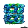
| Annotation | K12_GspD unpost-processing map | ||||||||||||
| Projections & Slices |
| ||||||||||||
| Density Histograms |
 Z
Z Y
Y X
X


- Sample components
-Entire : Full length T2SS secretin GspD complex
| Entire | Name: Full length T2SS secretin GspD complex |
|---|---|
| Components |
|
-Supramolecule #1: Full length T2SS secretin GspD complex
| Supramolecule | Name: Full length T2SS secretin GspD complex / type: complex / ID: 1 / Parent: 0 / Macromolecule list: all |
|---|---|
| Source (natural) | Organism: Escherichia coli K-12 (bacteria) |
| Molecular weight | Theoretical: 1 MDa |
-Supramolecule #2: Putative type II secretion system protein D
| Supramolecule | Name: Putative type II secretion system protein D / type: complex / ID: 2 / Parent: 1 / Macromolecule list: all |
|---|
-Macromolecule #1: Putative type II secretion system protein D
| Macromolecule | Name: Putative type II secretion system protein D / type: protein_or_peptide / ID: 1 / Number of copies: 15 / Enantiomer: LEVO |
|---|---|
| Source (natural) | Organism: Escherichia coli K-12 (bacteria) / Strain: K-12 |
| Molecular weight | Theoretical: 68.410789 KDa |
| Recombinant expression | Organism: Escherichia coli K-12 (bacteria) |
| Sequence | String: ENEQYGANFN NADIRQFVEI VGQHLGKTIL IDPSVQGTIS VRSNDTFSQQ EYYQFFLSIL DLYGYSVITL DNGFLKVVRS ANVKTSPGM IADSSRPGVG DELVTRIVPL ENVPARDLAP LLRQMMDAGS VGNVVHYEPS NVLILTGRAS TINKLIEVIK R VDVIGTEK ...String: ENEQYGANFN NADIRQFVEI VGQHLGKTIL IDPSVQGTIS VRSNDTFSQQ EYYQFFLSIL DLYGYSVITL DNGFLKVVRS ANVKTSPGM IADSSRPGVG DELVTRIVPL ENVPARDLAP LLRQMMDAGS VGNVVHYEPS NVLILTGRAS TINKLIEVIK R VDVIGTEK QQIIHLEYAS AEDLAEILNQ LISESHGKSQ MPALLSAKIV ADKRTNSLII SGPEKARQRI TSLLKSLDVE ES EEGNTRV YYLKYAKATN LVEVLTGVSE KLKDEKGNAR KPSSSGAMDN VAITADEQTN SLVITADQSV QEKLATVIAR LDI RRAQVL VEAIIVEVQD GNGLNLGVQW ANKNVGAQQF TNTGLPIFNA AQGVADYKKN GGITSANPAW DMFSAYNGMA AGFF NGDWG VLLTALASNN KNDILATPSI VTLDNKLASF NVGQDVPVLS GSQTTSGDNV FNTVERKTVG TKLKVTPQVN EGDAV LLEI EQEVSSVDSS SNSTLGPTFN TRTIQNAVLV KTGETVVLGG LLDDFSKEQV SKVPLLGDIP LVGQLFRYTS TERAKR NLM VFIRPTIIRD DDVYRSLSKE KYTRYRQEQQ QRIDGKSKAL VGSEDLPVLD ENTFNSHAPA PSSR UniProtKB: Putative secretin GspD |
-Experimental details
-Structure determination
| Method | cryo EM |
|---|---|
 Processing Processing | single particle reconstruction |
| Aggregation state | particle |
-Sample preparation
| Buffer | pH: 8 |
|---|---|
| Vitrification | Cryogen name: ETHANE |
- Electron microscopy
| Microscope | FEI TITAN KRIOS |
|---|---|
| Electron beam | Acceleration voltage: 300 kV / Electron source: FIELD EMISSION GUN |
| Electron optics | Illumination mode: OTHER / Imaging mode: BRIGHT FIELDBright-field microscopy / Cs: 2.7 mm |
| Sample stage | Specimen holder model: FEI TITAN KRIOS AUTOGRID HOLDER / Cooling holder cryogen: NITROGEN |
| Image recording | Film or detector model: GATAN K2 SUMMIT (4k x 4k) / Detector mode: SUPER-RESOLUTION / Average electron dose: 1.6 e/Å2 |
| Experimental equipment |  Model: Titan Krios / Image courtesy: FEI Company |
-Image processing
| Startup model | Type of model: OTHER |
|---|---|
| Initial angle assignment | Type: RANDOM ASSIGNMENT |
| Final angle assignment | Type: OTHER |
| Final reconstruction | Applied symmetry - Point group: C15 (15 fold cyclic) / Resolution.type: BY AUTHOR / Resolution: 3.04 Å / Resolution method: FSC 0.143 CUT-OFF / Software - Name: RELION / Number images used: 30659 |
-Atomic model buiding 1
| Refinement | Protocol: AB INITIO MODEL |
|---|---|
| Output model | PDB-5wq7: |
