 Movie
Movie Controller

[English] 日本語
 Yorodumi
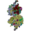
- EMDB-6664: The structure of rabbit skeletal muscle actomyosin rigor complex ... -
+ Open data

- Basic information
| Entry | Database: EMDB / ID: EMD-6664 | |||||||||
|---|---|---|---|---|---|---|---|---|---|---|
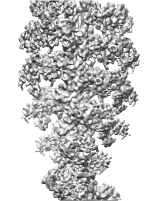
| Title | The structure of rabbit skeletal muscle actomyosin rigor complex at 5.2 angstrom. | |||||||||
 Map data Map data | ||||||||||
 Sample Sample |
| |||||||||
| Function / homology |  Function and homology information Function and homology information myosin complex / structural constituent of muscle / cytoskeletal motor activator activity / tropomyosin binding / myofibril / myosin heavy chain binding / mesenchyme migration / troponin I binding / actin filament bundle / filamentous actin ...myosin complex / structural constituent of muscle / cytoskeletal motor activator activity / tropomyosin binding / myofibril / myosin heavy chain binding / mesenchyme migration / troponin I binding / actin filament bundle / filamentous actin / actin filament bundle assembly / skeletal muscle thin filament assembly / cytoskeletal motor activity / striated muscle thin filament / skeletal muscle myofibril / actin monomer binding / skeletal muscle fiber development / stress fiber / titin binding / actin filament polymerization / cellular response to starvation / filopodium / actin filament / Hydrolases; Acting on acid anhydrides; Acting on acid anhydrides to facilitate cellular and subcellular movement / calcium-dependent protein binding / actin filament binding / lamellipodium / cell body / hydrolase activity / protein domain specific binding / calcium ion binding / positive regulation of gene expression / magnesium ion binding / ATP binding / identical protein binding / cytoplasm myosin complex / structural constituent of muscle / cytoskeletal motor activator activity / tropomyosin binding / myofibril / myosin heavy chain binding / mesenchyme migration / troponin I binding / actin filament bundle / filamentous actin ...myosin complex / structural constituent of muscle / cytoskeletal motor activator activity / tropomyosin binding / myofibril / myosin heavy chain binding / mesenchyme migration / troponin I binding / actin filament bundle / filamentous actin / actin filament bundle assembly / skeletal muscle thin filament assembly / cytoskeletal motor activity / striated muscle thin filament / skeletal muscle myofibril / actin monomer binding / skeletal muscle fiber development / stress fiber / titin binding / actin filament polymerization / cellular response to starvation / filopodium / actin filament / Hydrolases; Acting on acid anhydrides; Acting on acid anhydrides to facilitate cellular and subcellular movement / calcium-dependent protein binding / actin filament binding / lamellipodium / cell body / hydrolase activity / protein domain specific binding / calcium ion binding / positive regulation of gene expression / magnesium ion binding / ATP binding / identical protein binding / cytoplasmSimilarity search - Function | |||||||||
| Biological species |  Oryctolagus cuniculus (rabbit) / Rabbit (rabbit) Oryctolagus cuniculus (rabbit) / Rabbit (rabbit) | |||||||||
| Method | helical reconstruction / cryo EM / Resolution: 5.2 Å | |||||||||
 Authors Authors | Fujii T / Namba K | |||||||||
 Citation Citation | Journal: Nat Commun / Year: 2017 Title: Structure of actomyosin rigour complex at 5.2 Å resolution and insights into the ATPase cycle mechanism. Authors: Takashi Fujii / Keiichi Namba /  Abstract: Muscle contraction is driven by cyclic association and dissociation of myosin head of the thick filament with thin actin filament coupled with ATP binding and hydrolysis by myosin. However, because ...Muscle contraction is driven by cyclic association and dissociation of myosin head of the thick filament with thin actin filament coupled with ATP binding and hydrolysis by myosin. However, because of the absence of actomyosin rigour structure at high resolution, it still remains unclear how the strong binding of myosin to actin filament triggers the release of hydrolysis products and how ATP binding causes their dissociation. Here we report the structure of mammalian skeletal muscle actomyosin rigour complex at 5.2 Å resolution by electron cryomicroscopy. Comparison with the structures of myosin in various states shows a distinctly large conformational change, providing insights into the ATPase-coupled reaction cycle of actomyosin. Based on our observations, we hypothesize that asymmetric binding along the actin filament could function as a Brownian ratchet by favouring directionally biased thermal motions of myosin and actin. | |||||||||
| History |
|
- Structure visualization
| Movie |
Movie viewer |
|---|---|
| Structure viewer | EM map: SurfViewMolmilJmol/JSmol |
| Supplemental images |
- Downloads & links
-EMDB archive
| Map data | emd_6664.map.gz | 55.7 MB | EMDB map data format | |
|---|---|---|---|---|
| Header (meta data) | emd-6664-v30.xmlemd-6664.xml | 12.2 KB 12.2 KB | Display Display | EMDB header |
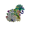
| Images |  emd_6664.png emd_6664.png | 71.5 KB | ||
| Archive directory |  http://ftp.pdbj.org/pub/emdb/structures/EMD-6664ftp://ftp.pdbj.org/pub/emdb/structures/EMD-6664 http://ftp.pdbj.org/pub/emdb/structures/EMD-6664ftp://ftp.pdbj.org/pub/emdb/structures/EMD-6664 | HTTPS FTP |
-Related structure data
| Related structure data |  5h53MC M: atomic model generated by this map C: citing same article ( |
|---|---|
| Similar structure data |

-Links
| EMDB pages | EMDB (EBI/PDBe) / EMDataResource |
|---|---|
| Related items in Molecule of the Month |


-Map
| File | Download / File: emd_6664.map.gz / Format: CCP4 / Size: 59.6 MB / Type: IMAGE STORED AS FLOATING POINT NUMBER (4 BYTES) | ||||||||||||||||||||||||||||||||||||||||||||||||||||||||||||||||||||
|---|---|---|---|---|---|---|---|---|---|---|---|---|---|---|---|---|---|---|---|---|---|---|---|---|---|---|---|---|---|---|---|---|---|---|---|---|---|---|---|---|---|---|---|---|---|---|---|---|---|---|---|---|---|---|---|---|---|---|---|---|---|---|---|---|---|---|---|---|---|
| Voxel size | X=Y=Z: 1.35 Å | ||||||||||||||||||||||||||||||||||||||||||||||||||||||||||||||||||||
| Density |
| ||||||||||||||||||||||||||||||||||||||||||||||||||||||||||||||||||||
| Symmetry | Space group: 1 | ||||||||||||||||||||||||||||||||||||||||||||||||||||||||||||||||||||
| Details | EMDB XML:
CCP4 map header:
| ||||||||||||||||||||||||||||||||||||||||||||||||||||||||||||||||||||
-Supplemental data
- Sample components
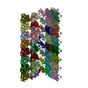
-Entire : Actomyosin rigor complex
| Entire | Name: Actomyosin rigor complex |
|---|---|
| Components |
|
-Supramolecule #1: Actomyosin rigor complex
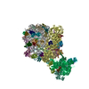
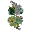
| Supramolecule | Name: Actomyosin rigor complex / type: complex / ID: 1 / Parent: 0 / Macromolecule list: #1-#4 |
|---|---|
| Source (natural) | Organism: Oryctolagus cuniculus (rabbit) |
-Macromolecule #1: Skeletal muscle myosin heavy chain MyHC-EO/IIL
| Macromolecule | Name: Skeletal muscle myosin heavy chain MyHC-EO/IIL / type: protein_or_peptide / ID: 1 / Number of copies: 1 / Enantiomer: LEVO |
|---|---|
| Source (natural) | Organism: Rabbit (rabbit) |
| Molecular weight | Theoretical: 96.770133 KDa |
| Sequence | String: MSSDAEMAIF GEAAPYLRKP EKERIEAQNR PFDSKKACFA VDDKEMYVKG MIQSRENDKV TVKTLDDRTL TLNSDQVFPM NPPKFDKIE DMAMMTHLHE PAVLYNLKER YAAWMIYTYS GLFCVTVNPY KWLPVYNPEV VTAYRGKKRQ EAPPHIFSIS D NAYQFMLT ...String: MSSDAEMAIF GEAAPYLRKP EKERIEAQNR PFDSKKACFA VDDKEMYVKG MIQSRENDKV TVKTLDDRTL TLNSDQVFPM NPPKFDKIE DMAMMTHLHE PAVLYNLKER YAAWMIYTYS GLFCVTVNPY KWLPVYNPEV VTAYRGKKRQ EAPPHIFSIS D NAYQFMLT DRDNQSILIT GESGAGKTVN TKRVIQYFAT IAVTGDKKKE QQPGKMQGTL EDQIIQANPL LEAFGNAKTV RN DNSSRFG KFIRIHFGAT GKLASADIET YLLEKSRVTF QLSSERSYHI FYQIMSNKKP ELIDLLLIST NPFDFPFVSQ GEV TVASID DSEELLATDN AIDILGFSSE EKVGIYKLTG AVMHYGNMKF KQKQREEQAE PDGTEVADKA GYLMGLNSAE MLKG LCCPR VKVGNEYVTK GQNVQQVTNS VGALAKAVYE KMFLWMVTRI NQQLDTKQPR QYFIGVLDIA GFEIFDFNSL EQLCI NFTN EKLQQFFNHH MFVLEQEEYK KEGIEWEFID FGMDLAACIE LIEKPMGIFS ILEEECMFPK ATDTSFKNKL YDQHLG KSN NFQKPKPAKG KAEAHFSLVH YAGTVDYNIA GWLDKNKDPL NETVVGLYQK SALKLLSFLF SNYAGAEAGD SGGSKKG GK KKGSSFQTVS AVFRENLNKL MTNLRSTHPH FVRCLIPNET KTPGVMDHYL VMHQLRCNGV LEGIRICRKG FPSRILYA D FKQRYRILNA SAIPEGQFID SKNASEKLLN SIDVDREQYR FGHTKVFFKA GLLGLLEEMR DEKLVTLMTR TQALCRGYL MRVEFKKMME RRDSIFCIQY NIRAFMNVKH WPWMNLFFKI KPLLK |
-Macromolecule #2: Myosin regulatory light chain 2, skeletal muscle isoform type 1
| Macromolecule | Name: Myosin regulatory light chain 2, skeletal muscle isoform type 1 type: protein_or_peptide / ID: 2 / Number of copies: 1 / Enantiomer: LEVO |
|---|---|
| Source (natural) | Organism: Rabbit (rabbit) |
| Molecular weight | Theoretical: 16.507588 KDa |
| Sequence | String: QTQIQEFKEA FTVIDQNRDG IIDKEDLRDT FAAMGRLNVK NEELDAMMKE ASGPINFTVF LTMFGEKLKG ADPEDVITGA FKVLDPEGK GTIKKQFLEE LLITQCDRFS QEEIKNMWAA FSPDVGGNVD YKNICYVITH GDAKDQE |
-Macromolecule #3: Myosin light chain 1/3, skeletal muscle isoform
| Macromolecule | Name: Myosin light chain 1/3, skeletal muscle isoform / type: protein_or_peptide / ID: 3 / Number of copies: 1 / Enantiomer: LEVO |
|---|---|
| Source (natural) | Organism: Rabbit (rabbit) |
| Molecular weight | Theoretical: 17.187293 KDa |
| Sequence | String: IKIEFSKEQQ DEFKEAFLLY DRTGDSKITL SQVGDVLRAL GTNPTNAEVK KVLGNPSNEE MNAKKIEFEQ FLPMLQAISN NKDQGTYED FVEGLRVFDK EGNGTVMGAE LRHVLATLGE KMKEEEVEAL MAGQEDSNGC INYEAFVKHI MSID |
-Macromolecule #4: Actin, alpha skeletal muscle
| Macromolecule | Name: Actin, alpha skeletal muscle / type: protein_or_peptide / ID: 4 / Number of copies: 2 / Enantiomer: LEVO |
|---|---|
| Source (natural) | Organism: Rabbit (rabbit) |
| Molecular weight | Theoretical: 41.875633 KDa |
| Sequence | String: DEDETTALVC DNGSGLVKAG FAGDDAPRAV FPSIVGRPRH QGVMVGMGQK DSYVGDEAQS KRGILTLKYP IE(HIC)GII TNW DDMEKIWHHT FYNELRVAPE EHPTLLTEAP LNPKANREKM TQIMFETFNV PAMYVAIQAV LSLYASGRTT GIVLDSG DG VTHNVPIYEG ...String: DEDETTALVC DNGSGLVKAG FAGDDAPRAV FPSIVGRPRH QGVMVGMGQK DSYVGDEAQS KRGILTLKYP IE(HIC)GII TNW DDMEKIWHHT FYNELRVAPE EHPTLLTEAP LNPKANREKM TQIMFETFNV PAMYVAIQAV LSLYASGRTT GIVLDSG DG VTHNVPIYEG YALPHAIMRL DLAGRDLTDY LMKILTERGY SFVTTAEREI VRDIKEKLCY VALDFENEMA TAASSSSL E KSYELPDGQV ITIGNERFRC PETLFQPSFI GMESAGIHET TYNSIMKCDI DIRKDLYANN VMSGGTTMYP GIADRMQKE ITALAPSTMK IKIIAPPERK YSVWIGGSIL ASLSTFQQMW ITKQEYDEAG PSIVHRKCF |
-Macromolecule #5: ADENOSINE-5'-DIPHOSPHATE
| Macromolecule | Name: ADENOSINE-5'-DIPHOSPHATE / type: ligand / ID: 5 / Number of copies: 2 / Formula: ADP |
|---|---|
| Molecular weight | Theoretical: 427.201 Da |
| Chemical component information |  ChemComp-ADP: |
-Experimental details
-Structure determination
| Method | cryo EM |
|---|---|
 Processing Processing | helical reconstruction |
| Aggregation state | filament |
-Sample preparation
| Buffer | pH: 7.5 |
|---|---|
| Vitrification | Cryogen name: ETHANE |
- Electron microscopy
| Microscope | JEOL 3200FSC |
|---|---|
| Electron beam | Acceleration voltage: 200 kV / Electron source: FIELD EMISSION GUN |
| Electron optics | Illumination mode: FLOOD BEAM / Imaging mode: BRIGHT FIELDBright-field microscopy |
| Image recording | Film or detector model: TVIPS TEMCAM-F415 (4k x 4k) / Average electron dose: 20.0 e/Å2 |
-Image processing
| Final angle assignment | Type: NOT APPLICABLE |
|---|---|
| Final reconstruction | Applied symmetry - Helical parameters - Δz: 27.6 Å Applied symmetry - Helical parameters - Δ&Phi: 166.7 ° Applied symmetry - Helical parameters - Axial symmetry: C1 (asymmetric) Resolution.type: BY AUTHOR / Resolution: 5.2 Å / Resolution method: FSC 0.143 CUT-OFF / Number images used: 31535 |
