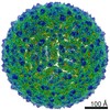
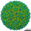
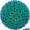
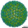
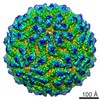
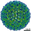
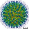
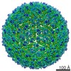
Journal: Nat Struct Mol Biol / Year: 2013 Title: Cryo-EM structure of the mature dengue virus at 3.5-Å resolution. Authors: Xiaokang Zhang / Peng Ge / Xuekui Yu / Jennifer M Brannan / Guoqiang Bi / Qinfen Zhang / Stan Schein / Z Hong Zhou / Abstract: Regulated by pH, membrane-anchored proteins E and M function during dengue virus maturation and membrane fusion. Our atomic model of the whole virion from cryo-electron microscopy at 3.5-Å ...Regulated by pH, membrane-anchored proteins E and M function during dengue virus maturation and membrane fusion. Our atomic model of the whole virion from cryo-electron microscopy at 3.5-Å resolution reveals that in the mature virus at neutral extracellular pH, the N-terminal 20-amino-acid segment of M (involving three pH-sensing histidines) latches and thereby prevents spring-loaded E fusion protein from prematurely exposing its fusion peptide. This M latch is fastened at an earlier stage, during maturation at acidic pH in the trans-Golgi network. At a later stage, to initiate infection in response to acidic pH in the late endosome, M releases the latch and exposes the fusion peptide. Thus, M serves as a multistep chaperone of E to control the conformational changes accompanying maturation and infection. These pH-sensitive interactions could serve as targets for drug discovery.
History
Deposition
Oct 10, 2012
-
Header (metadata) release
Nov 28, 2012
-
Map release
Dec 19, 2012
-
Update
Jan 16, 2013
-
Current status
Jan 16, 2013
Processing site: RCSB / Status: Released
-
Structure visualization
Movie
Surface view with section colored by density value
Category: FILM / Film or detector model: KODAK SO-163 FILM / Digitization - Scanner: NIKON SUPER COOLSCAN 9000 / Digitization - Sampling interval: 6.35 µm / Number real images: 1103 / Average electron dose: 25 e/Å2
Experimental equipment
Model: Titan Krios / Image courtesy: FEI Company
-
Image processing
CTF correction
Details: EMAN per particle, with astigmatism compensation
Final reconstruction
Algorithm: OTHER / Resolution.type: BY AUTHOR / Resolution: 3.6 Å / Resolution method: FSC 0.143 CUT-OFF / Software - Name: EMAN / Number images used: 9288
Details
EMAN with Multi-path Simulated Annealing
+
About Yorodumi
-
News
-
Feb 9, 2022. New format data for meta-information of EMDB entries
New format data for meta-information of EMDB entries
Version 3 of the EMDB header file is now the official format.
The previous official version 1.9 will be removed from the archive.
In the structure databanks used in Yorodumi, some data are registered as the other names, "COVID-19 virus" and "2019-nCoV". Here are the details of the virus and the list of structure data.
Jan 31, 2019. EMDB accession codes are about to change! (news from PDBe EMDB page)
EMDB accession codes are about to change! (news from PDBe EMDB page)
The allocation of 4 digits for EMDB accession codes will soon come to an end. Whilst these codes will remain in use, new EMDB accession codes will include an additional digit and will expand incrementally as the available range of codes is exhausted. The current 4-digit format prefixed with “EMD-” (i.e. EMD-XXXX) will advance to a 5-digit format (i.e. EMD-XXXXX), and so on. It is currently estimated that the 4-digit codes will be depleted around Spring 2019, at which point the 5-digit format will come into force.
The EM Navigator/Yorodumi systems omit the EMD- prefix.
Related info.:Q: What is EMD? / ID/Accession-code notation in Yorodumi/EM Navigator
Yorodumi is a browser for structure data from EMDB, PDB, SASBDB, etc.
This page is also the successor to EM Navigator detail page, and also detail information page/front-end page for Omokage search.
The word "yorodu" (or yorozu) is an old Japanese word meaning "ten thousand". "mi" (miru) is to see.
Related info.:EMDB / PDB / SASBDB / Comparison of 3 databanks / Yorodumi Search / Aug 31, 2016. New EM Navigator & Yorodumi / Yorodumi Papers / Jmol/JSmol / Function and homology information / Changes in new EM Navigator and Yorodumi
 Movie
Movie Controller


 Open data
 Basic information
 Map data
 Sample
 Keywords
 flavivirus /
  Function and homology information
 Authors
 Citation
 Structure visualization
 Downloads & links
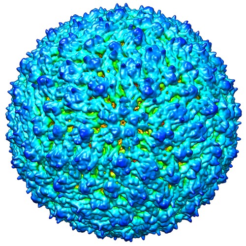 emd_5520_1.jpg
 http://ftp.pdbj.org/pub/emdb/structures/EMD-5520


 Sample components
 Processing
 Electron microscopy
