 Movie
Movie Controller

[English] 日本語
 Yorodumi
- EMDB-5389: An RNA Degradation Machine Sculpted by Ro Autoantigen and Noncodi... -
+ Open data

- Basic information
| Entry | Database: EMDB / ID: EMD-5389 | |||||||||
|---|---|---|---|---|---|---|---|---|---|---|
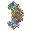
| Title | An RNA Degradation Machine Sculpted by Ro Autoantigen and Noncoding RNA | |||||||||
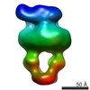
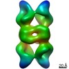
 Map data Map data | negative stain single particle reconstruction of GraFix-prepared Rsr/Y RNA/PNPase complex | |||||||||
 Sample Sample |
| |||||||||
 Keywords Keywords |  exonuclease / Y RNA / degradation machine exonuclease / Y RNA / degradation machine | |||||||||
| Biological species |  Deinococcus radiodurans (radioresistant) Deinococcus radiodurans (radioresistant) | |||||||||
| Method | single particle reconstruction / negative staining / Resolution: 25.0 Å | |||||||||
 Authors Authors | Chen X / Taylor DW / Wang HW / Wolin SL | |||||||||
 Citation Citation | Journal: Cell / Year: 2013 Title: An RNA degradation machine sculpted by Ro autoantigen and noncoding RNA. Authors: Xinguo Chen / David W Taylor / Casey C Fowler / Jorge E Galan / Hong-Wei Wang / Sandra L Wolin /  Abstract: Many bacteria contain an ortholog of the Ro autoantigen, a ring-shaped protein that binds noncoding RNAs (ncRNAs) called Y RNAs. In the only studied bacterium, Deinococcus radiodurans, the Ro ...Many bacteria contain an ortholog of the Ro autoantigen, a ring-shaped protein that binds noncoding RNAs (ncRNAs) called Y RNAs. In the only studied bacterium, Deinococcus radiodurans, the Ro ortholog Rsr functions in heat-stress-induced ribosomal RNA (rRNA) maturation and starvation-induced rRNA decay. However, the mechanism by which this conserved protein and its associated ncRNAs act has been obscure. We report that Rsr and the exoribonuclease polynucleotide phosphorylase (PNPase) form an RNA degradation machine that is scaffolded by Y RNA. Single-particle electron microscopy, followed by docking of atomic models into the reconstruction, suggests that Rsr channels single-stranded RNA into the PNPase cavity. Biochemical assays reveal that Rsr and Y RNA adapt PNPase for effective degradation of structured RNAs. A Ro ortholog and ncRNA also associate with PNPase in Salmonella Typhimurium. Our studies identify another ribonucleoprotein machine and demonstrate that ncRNA, by tethering a protein cofactor, can alter the substrate specificity of an enzyme. | |||||||||
| History |
|
- Structure visualization
| Movie |
 Movie viewer Movie viewer |
|---|---|
| Structure viewer | EM map: SurfViewMolmilJmol/JSmol |
| Supplemental images |

- Downloads & links
-EMDB archive
| Map data | emd_5389.map.gz | 1.3 MB | EMDB map data format | |
|---|---|---|---|---|
| Header (meta data) | emd-5389-v30.xmlemd-5389.xml | 14.3 KB 14.3 KB | Display Display | EMDB header |
| Images | 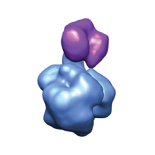 emd_5389_1.png emd_5389_1.png | 75.2 KB | ||
| Archive directory |  http://ftp.pdbj.org/pub/emdb/structures/EMD-5389ftp://ftp.pdbj.org/pub/emdb/structures/EMD-5389 http://ftp.pdbj.org/pub/emdb/structures/EMD-5389ftp://ftp.pdbj.org/pub/emdb/structures/EMD-5389 | HTTPS FTP |
-Related structure data
| Similar structure data |
|---|


-Links
| EMDB pages | EMDB (EBI/PDBe) / EMDataResource |
|---|
-Map
| File | Download / File: emd_5389.map.gz / Format: CCP4 / Size: 1.4 MB / Type: IMAGE STORED AS FLOATING POINT NUMBER (4 BYTES) | ||||||||||||||||||||||||||||||||||||||||||||||||||||||||||||||||||||
|---|---|---|---|---|---|---|---|---|---|---|---|---|---|---|---|---|---|---|---|---|---|---|---|---|---|---|---|---|---|---|---|---|---|---|---|---|---|---|---|---|---|---|---|---|---|---|---|---|---|---|---|---|---|---|---|---|---|---|---|---|---|---|---|---|---|---|---|---|---|
| Annotation | negative stain single particle reconstruction of GraFix-prepared Rsr/Y RNA/PNPase complex | ||||||||||||||||||||||||||||||||||||||||||||||||||||||||||||||||||||
| Voxel size | X=Y=Z: 4.36 Å | ||||||||||||||||||||||||||||||||||||||||||||||||||||||||||||||||||||
| Density |
| ||||||||||||||||||||||||||||||||||||||||||||||||||||||||||||||||||||
| Symmetry | Space group: 1 | ||||||||||||||||||||||||||||||||||||||||||||||||||||||||||||||||||||
| Details | EMDB XML:
CCP4 map header:
| ||||||||||||||||||||||||||||||||||||||||||||||||||||||||||||||||||||
-Supplemental data
- Sample components
-Entire : Rsr/Y RNA/PNPase complex
| Entire | Name: Rsr/Y RNA/PNPase complex |
|---|---|
| Components |
|
-Supramolecule #1000: Rsr/Y RNA/PNPase complex
| Supramolecule | Name: Rsr/Y RNA/PNPase complex / type: sample / ID: 1000 / Details: The sample was monodisperse and homogeneous. Oligomeric state: one Rsr-Y RNA complex binds to one PNPase trimer Number unique components: 3 |
|---|---|
| Molecular weight | Experimental: 375 KDa / Theoretical: 365 KDa / Method: glycerol gradient |
-Macromolecule #1: Ro sixty-related
| Macromolecule | Name: Ro sixty-related / type: protein_or_peptide / ID: 1 / Name.synonym: Rsr, Ro 60 kDa autoantigen / Details: His-tag Rsr / Number of copies: 1 / Oligomeric state: monomer / Recombinant expression: Yes |
|---|---|
| Source (natural) | Organism: Deinococcus radiodurans (radioresistant) |
| Molecular weight | Theoretical: 62 KDa |
| Recombinant expression | Organism: Escherichia coli (E. coli) / Recombinant plasmid: RSFDuet-1 |
-Macromolecule #3: polynucleotide phosphorylase
| Macromolecule | Name: polynucleotide phosphorylase / type: protein_or_peptide / ID: 3 / Name.synonym: PNPase / Details: Strep-tag PNPase / Number of copies: 3 / Oligomeric state: Trimer / Recombinant expression: Yes |
|---|---|
| Source (natural) | Organism: Deinococcus radiodurans (radioresistant) |
| Molecular weight | Theoretical: 262 KDa |
| Recombinant expression | Organism: Escherichia coli (E. coli) / Recombinant plasmid: RSFDuet-1 |
-Macromolecule #2: Y RNA
| Macromolecule | Name: Y RNA / type: rna / ID: 2 Details: Y RNA generated with correct 3' end using hammerhead ribozyme Classification: OTHER / Structure: OTHER / Synthetic?: No |
|---|---|
| Source (natural) | Organism: Deinococcus radiodurans (radioresistant) |
| Molecular weight | Theoretical: 42 KDa |
-Experimental details
-Structure determination
| Method | negative staining |
|---|---|
 Processing Processing | single particle reconstruction |
| Aggregation state | particle |
-Sample preparation
| Concentration | 0.1 mg/mL |
|---|---|
| Buffer | pH: 7.5 Details: 20 mM HEPES, 50 mM NaCl, 2 mM beta-mercaptoethanol, 1 mM MgCl2, 1 mM MnCl2, 1 mM Petabloc |
| Staining | Type: NEGATIVE Details: Grids with adsorbed protein were stained consecutively with 3 droplets of 2% w/v uranyl acetate for 10 seconds each. |
| Grid | Details: Homemade holey carbon grids with a thin layer of carbon over the holes |
| Vitrification | Cryogen name: NONE / Instrument: OTHER |
- Electron microscopy
| Microscope | FEI TECNAI 12 |
|---|---|
| Electron beam | Acceleration voltage: 120 kV / Electron source: LAB6 |
| Electron optics | Illumination mode: FLOOD BEAM / Imaging mode: BRIGHT FIELDBright-field microscopy / Cs: 2.3 mm / Nominal defocus max: 1.2 µm / Nominal defocus min: 0.8 µm / Nominal magnification: 49000 |
| Sample stage | Specimen holder model: OTHER |
| Alignment procedure | Legacy - Astigmatism: object lens astigmatism was corrected at 42,000 magnification |
| Date | Jul 12, 2009 |
| Image recording | Category: CCD / Film or detector model: TVIPS TEMCAM-F416 (4k x 4k) / Number real images: 40 / Average electron dose: 20 e/Å2 |
-Image processing
| CTF correction | Details: Each particle in IMAGIC |
|---|---|
| Final two d classification | Number classes: 50 |
| Final reconstruction | Algorithm: OTHER / Resolution.type: BY AUTHOR / Resolution: 25.0 Å / Resolution method: FSC 0.5 CUT-OFF / Software - Name: LEGINON, EMAN2, SPARX / Number images used: 9000 |
| Details | The particles were selected manually using boxer in EMAN. |
-Atomic model buiding 1
| Initial model | PDB ID: Chain - Chain ID: A |
|---|---|
| Software | Name: Chimera |
| Details | Protocol: Rigid body using Fit-in-Map. A trimer of the crystal structure of PNPase was created using COOT and used for fitting. |
| Refinement | Space: REAL / Protocol: RIGID BODY FIT / Target criteria: Cross-correlation coefficient |

-Atomic model buiding 2
| Initial model | PDB ID: Chain - #0 - Chain ID: B / Chain - #1 - Chain ID: E / Chain - #2 - Chain ID: F / Chain - #3 - Chain ID: H |
|---|---|
| Software | Name: Chimera |
| Details | Protocol: Rigid body using Fit-in-Map. Combined with the double-stranded portion of the misfolded substrate RNA from PDB 2I91. |
| Refinement | Space: REAL / Protocol: RIGID BODY FIT / Target criteria: Cross-correlation coefficient |
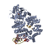
-Atomic model buiding 3
| Initial model | PDB ID: Chain - #0 - Chain ID: C / Chain - #1 - Chain ID: D |
|---|---|
| Software | Name: Chimera |
| Details | Protocol: Rigid body using Fit-in-Map. The double-stranded portion of the misfolded substrate RNA from this structure was combined with PDB 1YVP to create a model of Rsr bound by Y RNA and a full substrate RNA with single-stranded tail. |
| Refinement | Space: REAL / Protocol: RIGID BODY FIT / Target criteria: Cross-correlation coefficient |

