 Movie
Movie Controller

[English] 日本語
 Yorodumi
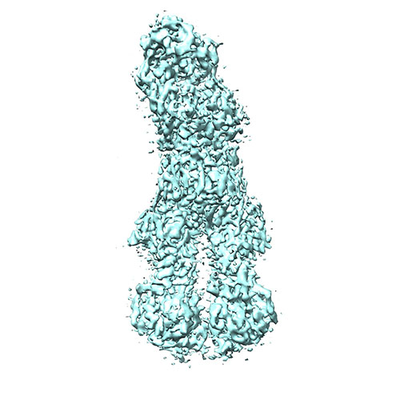
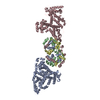
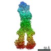
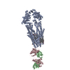
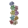
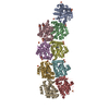
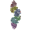
- EMDB-4282: Apo form of UIC2 Fab complex of human-mouse chimeric ABCB1 (ABCB1HM) -
+ Open data

- Basic information
| Entry | Database: EMDB / ID: EMD-4282 | ||||||||||||
|---|---|---|---|---|---|---|---|---|---|---|---|---|---|
| Title | Apo form of UIC2 Fab complex of human-mouse chimeric ABCB1 (ABCB1HM) | ||||||||||||
 Map data Map data | Human-mouse chimeric ABCB1 (ABCB1Hm) complex with UIC2 Antigen binding fragment | ||||||||||||
 Sample Sample |
| ||||||||||||
| Function / homology |  Function and homology information Function and homology informationpositive regulation of anion channel activity / ceramide translocation / terpenoid transport / ceramide floppase activity / carboxylic acid transmembrane transport / carboxylic acid transmembrane transporter activity /  floppase activity / Abacavir transmembrane transport / external side of apical plasma membrane / regulation of response to osmotic stress ...positive regulation of anion channel activity / ceramide translocation / terpenoid transport / ceramide floppase activity / carboxylic acid transmembrane transport / carboxylic acid transmembrane transporter activity / floppase activity / Abacavir transmembrane transport / external side of apical plasma membrane / regulation of response to osmotic stress / Atorvastatin ADME / phosphatidylcholine floppase activity / phosphatidylethanolamine flippase activity / xenobiotic transport across blood-brain barrier / xenobiotic detoxification by transmembrane export across the plasma membrane / export across plasma membrane / ABC-type xenobiotic transporter / transepithelial transport / P-type phospholipid transporter / ABC-type xenobiotic transporter activity / phospholipid translocation / Prednisone ADME / xenobiotic transmembrane transporter activity / efflux transmembrane transporter activity / transmembrane transporter activity / transport across blood-brain barrier / ATPase-coupled transmembrane transporter activity / xenobiotic metabolic process / regulation of chloride transport / stem cell proliferation / ABC-family proteins mediated transport / transmembrane transport / G2/M transition of mitotic cell cycle / response to xenobiotic stimulus / apical plasma membrane / ubiquitin protein ligase binding / cell surface / ATP hydrolysis activity / extracellular exosome / ATP binding / membrane / plasma membrane / cytoplasm floppase activity / Abacavir transmembrane transport / external side of apical plasma membrane / regulation of response to osmotic stress ...positive regulation of anion channel activity / ceramide translocation / terpenoid transport / ceramide floppase activity / carboxylic acid transmembrane transport / carboxylic acid transmembrane transporter activity / floppase activity / Abacavir transmembrane transport / external side of apical plasma membrane / regulation of response to osmotic stress / Atorvastatin ADME / phosphatidylcholine floppase activity / phosphatidylethanolamine flippase activity / xenobiotic transport across blood-brain barrier / xenobiotic detoxification by transmembrane export across the plasma membrane / export across plasma membrane / ABC-type xenobiotic transporter / transepithelial transport / P-type phospholipid transporter / ABC-type xenobiotic transporter activity / phospholipid translocation / Prednisone ADME / xenobiotic transmembrane transporter activity / efflux transmembrane transporter activity / transmembrane transporter activity / transport across blood-brain barrier / ATPase-coupled transmembrane transporter activity / xenobiotic metabolic process / regulation of chloride transport / stem cell proliferation / ABC-family proteins mediated transport / transmembrane transport / G2/M transition of mitotic cell cycle / response to xenobiotic stimulus / apical plasma membrane / ubiquitin protein ligase binding / cell surface / ATP hydrolysis activity / extracellular exosome / ATP binding / membrane / plasma membrane / cytoplasmSimilarity search - Function | ||||||||||||
| Biological species |  Homo sapiens (human) / Homo sapiens (human) /  Mus musculus (house mouse) Mus musculus (house mouse) | ||||||||||||
| Method | single particle reconstruction / cryo EM / Resolution: 4.14 Å | ||||||||||||
 Authors Authors | Alam A / Locher KP | ||||||||||||
| Funding support |  Switzerland, Switzerland,  United States, 3 items United States, 3 items
| ||||||||||||
 Citation Citation | Journal: Proc Natl Acad Sci U S A / Year: 2018 Title: Structure of a zosuquidar and UIC2-bound human-mouse chimeric ABCB1. Authors: Amer Alam / Raphael Küng / Julia Kowal / Robert A McLeod / Nina Tremp / Eugenia V Broude / Igor B Roninson / Henning Stahlberg / Kaspar P Locher / Abstract: The multidrug transporter ABCB1 (P-glycoprotein) is an ATP-binding cassette transporter that has a key role in protecting tissues from toxic insult and contributes to multidrug extrusion from cancer ...The multidrug transporter ABCB1 (P-glycoprotein) is an ATP-binding cassette transporter that has a key role in protecting tissues from toxic insult and contributes to multidrug extrusion from cancer cells. Here, we report the near-atomic resolution cryo-EM structure of nucleotide-free ABCB1 trapped by an engineered disulfide cross-link between the nucleotide-binding domains (NBDs) and bound to the antigen-binding fragment of the human-specific inhibitory antibody UIC2 and to the third-generation ABCB1 inhibitor zosuquidar. Our structure reveals the transporter in an occluded conformation with a central, enclosed, inhibitor-binding pocket lined by residues from all transmembrane (TM) helices of ABCB1. The pocket spans almost the entire width of the lipid membrane and is occupied exclusively by two closely interacting zosuquidar molecules. The external, conformational epitope facilitating UIC2 binding is also visualized, providing a basis for its inhibition of substrate efflux. Additional cryo-EM structures suggest concerted movement of TM helices from both halves of the transporters associated with closing the NBD gap, as well as zosuquidar binding. Our results define distinct recognition interfaces of ABCB1 inhibitory agents, which may be exploited for therapeutic purposes. | ||||||||||||
| History |
|
- Structure visualization
| Movie |
Movie viewer |
|---|---|
| Structure viewer | EM map: SurfViewMolmilJmol/JSmol |
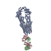
| Supplemental images |
- Downloads & links
-EMDB archive
| Map data | emd_4282.map.gz | 28.6 MB | EMDB map data format | |
|---|---|---|---|---|
| Header (meta data) | emd-4282-v30.xmlemd-4282.xml | 16.1 KB 16.1 KB | Display Display | EMDB header |
| Images |  emd_4282.png emd_4282.png | 124.6 KB | ||
| Archive directory |  http://ftp.pdbj.org/pub/emdb/structures/EMD-4282ftp://ftp.pdbj.org/pub/emdb/structures/EMD-4282 http://ftp.pdbj.org/pub/emdb/structures/EMD-4282ftp://ftp.pdbj.org/pub/emdb/structures/EMD-4282 | HTTPS FTP |
-Related structure data
| Related structure data |  6fn4MC 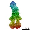 4281C 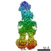 4283C 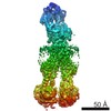 4284C 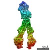 4285C 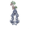 6fn1C C: citing same article ( M: atomic model generated by this map |
|---|---|
| Similar structure data |
-Links
| EMDB pages | EMDB (EBI/PDBe) / EMDataResource |
|---|---|
| Related items in Molecule of the Month |


-Map
| File | Download / File: emd_4282.map.gz / Format: CCP4 / Size: 30.5 MB / Type: IMAGE STORED AS FLOATING POINT NUMBER (4 BYTES) | ||||||||||||||||||||||||||||||||||||||||||||||||||||||||||||
|---|---|---|---|---|---|---|---|---|---|---|---|---|---|---|---|---|---|---|---|---|---|---|---|---|---|---|---|---|---|---|---|---|---|---|---|---|---|---|---|---|---|---|---|---|---|---|---|---|---|---|---|---|---|---|---|---|---|---|---|---|---|
| Annotation | Human-mouse chimeric ABCB1 (ABCB1Hm) complex with UIC2 Antigen binding fragment | ||||||||||||||||||||||||||||||||||||||||||||||||||||||||||||
| Voxel size | X=Y=Z: 1.387 Å | ||||||||||||||||||||||||||||||||||||||||||||||||||||||||||||
| Density |
| ||||||||||||||||||||||||||||||||||||||||||||||||||||||||||||
| Symmetry | Space group: 1 | ||||||||||||||||||||||||||||||||||||||||||||||||||||||||||||
| Details | EMDB XML:
CCP4 map header:
| ||||||||||||||||||||||||||||||||||||||||||||||||||||||||||||
-Supplemental data
- Sample components
-Entire : Apo Human-mouse chimeric ABCB1 (ABCB1HM) complex with UIC2 fab
| Entire | Name: Apo Human-mouse chimeric ABCB1 (ABCB1HM) complex with UIC2 fab |
|---|---|
| Components |
|
-Supramolecule #1: Apo Human-mouse chimeric ABCB1 (ABCB1HM) complex with UIC2 fab
| Supramolecule | Name: Apo Human-mouse chimeric ABCB1 (ABCB1HM) complex with UIC2 fab type: complex / ID: 1 / Parent: 0 / Macromolecule list: #1-#3 |
|---|---|
| Molecular weight | Theoretical: 195 KDa |
-Supramolecule #2: Apo Human-mouse chimeric ABCB1 (ABCB1HM) complex with UIC2 fab
| Supramolecule | Name: Apo Human-mouse chimeric ABCB1 (ABCB1HM) complex with UIC2 fab type: complex / ID: 2 / Parent: 1 / Macromolecule list: #1 |
|---|---|
| Source (natural) | Organism: Homo sapiens (human) |
| Recombinant expression | Organism: Homo sapiens (human) |
| Molecular weight | Theoretical: 142 KDa |
-Supramolecule #3: Apo Human-mouse chimeric ABCB1 (ABCB1HM) complex with UIC2 fab
| Supramolecule | Name: Apo Human-mouse chimeric ABCB1 (ABCB1HM) complex with UIC2 fab type: complex / ID: 3 / Parent: 1 / Macromolecule list: #2-#3 |
|---|---|
| Source (natural) | Organism: Mus musculus (house mouse) |
| Recombinant expression | Organism: Mus musculus (house mouse) |
| Molecular weight | Theoretical: 48.7 KDa |
-Macromolecule #1: Apo form of Human-mouse chimeric ABCB1 (ABCB1HM) in complex with ...
| Macromolecule | Name: Apo form of Human-mouse chimeric ABCB1 (ABCB1HM) in complex with Antigen binding fragment of UIC2. type: protein_or_peptide / ID: 1 / Number of copies: 1 / Enantiomer: LEVO |
|---|---|
| Source (natural) | Organism: Homo sapiens (human) |
| Molecular weight | Theoretical: 141.283156 KDa |
| Recombinant expression | Organism: Homo sapiens (human) |
| Sequence | String: MELEEDLKGR ADKNFSKMGK KSKKEKKEKK PAVSVLTMFR YAGWLDRLYM LVGTLAAIIH GVALPLMMLI FGEMTDIFAN AGNLEDLMS NITNRSDIND TGFFMNLEED MTTYAYYYTG IGAGVLIVAY IQVSFWLLAA GRQIHKIRQK FFHAIMNQEI G WFDVHDVG ...String: MELEEDLKGR ADKNFSKMGK KSKKEKKEKK PAVSVLTMFR YAGWLDRLYM LVGTLAAIIH GVALPLMMLI FGEMTDIFAN AGNLEDLMS NITNRSDIND TGFFMNLEED MTTYAYYYTG IGAGVLIVAY IQVSFWLLAA GRQIHKIRQK FFHAIMNQEI G WFDVHDVG ELNTRLTDDV SKINEGIGDK IGMFFQAMAT FFGGFIIGFT RGWKLTLVIL AISPVLGLSA GIWAKILSSF TD KELHAYA KAGAVAEEVL AAIRTVIAFG GQKKELERYN NNLEEAKRLG IKKAITANIS MGAAFLLIYA SYALAFWYGT TLV LSGEYS IGQVLTVFFS VLIGAFSVGQ ASPNIEAFAN ARGAAYEVFK IIDNKPSIDS FSKSGHKPDN IQGNLEFKNI HFSY PSRKE VQILKGLNLK VKSGQTVALV GNSGAGKSTT VQLMQRLYDP LDGMVSIDGQ DIRTINVRYL REIIGVVSQE PVLFA TTIA ENIRYGREDV TMDEIEKAVK EANAYDFIMK LPHQFDTLVG ERGAQLSGGQ KQRIAIARAL VRNPKILLLD EATCAL DTE SEAVVQAALD KAREGRTTIV IAHRLSTVRN ADVIAGFDGG VIVEQGNHDE LMREKGIYFK LVMTQTAGNE IELGNEA AK SKDEIDNLDM SSKDSGSSLI RRRSTRKSIA GPHDQDRKLS TKEALDEDVP PASFWRILKL NSTEWPYFVV GIFVAIIN G GLQPAFSVIF SKIIGVFTRI DDPETKRQNS NLFSLLFLIL GIISFITFFL QGFTFGKAGE ILTKRLRYMV FKSMLRQDV SWFDDPKNTT GALTTRLAND AAQVKGATGS RLAVIFQNIA NLGTGIIISF IYGWQLTLLL LAIVPIIAIA GVVEMKMLSG QALKDKKEL EGSGKIATEA IENFRTVVSL TREQKFETMY AQSLQIPYRN AMKKAHVFGI TFSFTQAMMY FSYAAAFRFG A YLVAHKLM SFEDVLLVFS AIVFGAMAVG QVSSFAPDYA KATVSASHII RIIEKTPEID SYSTQGLKPN MLEGNVQFSG VV FNYPTRP SIPVLQGLSL EVKKGQTLAL VGSSGAGKST VVQLLERFYD PMAGSVFLDG KEIKQLNVQW LRAQLGIVSQ EPI LFDTSI AENIAYGDNS RVVSYEEIVR AAKEANIHQF IDSLPDKYNT RVGDKGTQLS GGQKQRIAIA RALVRQPHIL LLDE ATCAL DTESEKVVQE ALDKAREGRT TIVIAHRLST IQNADLIVVI QNGKVKEHGT HQQLLAQKGI YFSMVSVQAG AKRS |
-Macromolecule #2: UIC2 Antigen Binding Fragment Light chain
| Macromolecule | Name: UIC2 Antigen Binding Fragment Light chain / type: protein_or_peptide / ID: 2 / Number of copies: 1 / Enantiomer: LEVO |
|---|---|
| Source (natural) | Organism: Mus musculus (house mouse) |
| Molecular weight | Theoretical: 24.321039 KDa |
| Recombinant expression | Organism: Mus musculus (house mouse) |
| Sequence | String: QVVMTQSPLS LPVSLGDQAS ISCRSSQSLL HSNGNTYLHW YLQKPGQSPK LLIYKVSNRF SGVPDRFSGS GSGTDFTLKI SRVEAEDLG VYFCSQSTHI PPWTFGGGTK LDIKRADAAP TVSIFPPSSE QLTSGGLSVV CFLNNFYPKD INVKWKIDGS E RQNGVLNS ...String: QVVMTQSPLS LPVSLGDQAS ISCRSSQSLL HSNGNTYLHW YLQKPGQSPK LLIYKVSNRF SGVPDRFSGS GSGTDFTLKI SRVEAEDLG VYFCSQSTHI PPWTFGGGTK LDIKRADAAP TVSIFPPSSE QLTSGGLSVV CFLNNFYPKD INVKWKIDGS E RQNGVLNS WTDQDSKDST YSMSSTLTLT KDEYERHNSY TCEATHKTST SPIVKSFNRN EC |
-Macromolecule #3: UIC2 Antigen binding fragment Heavy chain
| Macromolecule | Name: UIC2 Antigen binding fragment Heavy chain / type: protein_or_peptide / ID: 3 / Number of copies: 1 / Enantiomer: LEVO |
|---|---|
| Source (natural) | Organism: Mus musculus (house mouse) |
| Molecular weight | Theoretical: 24.381281 KDa |
| Recombinant expression | Organism: Mus musculus (house mouse) |
| Sequence | String: EVQLQESGPE LVKTGASVKI SCKASGYSFS NYYIHWVKQS HGKSLEWIGF ISCYNGATFY NQKFKGKATF TVDNSSSTAY MKFNSLTFE DSAVYYCARL PIQFGNFYPM DYWGQGTTVT VSSAKTTAPS VYPLAPVCGD TTGSSVTLGC LVKGYFPEPV T LTWNSGSL ...String: EVQLQESGPE LVKTGASVKI SCKASGYSFS NYYIHWVKQS HGKSLEWIGF ISCYNGATFY NQKFKGKATF TVDNSSSTAY MKFNSLTFE DSAVYYCARL PIQFGNFYPM DYWGQGTTVT VSSAKTTAPS VYPLAPVCGD TTGSSVTLGC LVKGYFPEPV T LTWNSGSL SSGVHTFPAV LQSDLYTLSS SVTVTSSTWP SQSITCNVAH PASSTKVDKK IEPRGPT |
-Macromolecule #4: 2-acetamido-2-deoxy-beta-D-glucopyranose
| Macromolecule | Name: 2-acetamido-2-deoxy-beta-D-glucopyranose / type: ligand / ID: 4 / Number of copies: 3 / Formula: NAG |
|---|---|
| Molecular weight | Theoretical: 221.208 Da |
| Chemical component information |  ChemComp-NAG: |
-Macromolecule #5: 1,2-DIACYL-SN-GLYCERO-3-PHOSPHOETHANOLAMINE
| Macromolecule | Name: 1,2-DIACYL-SN-GLYCERO-3-PHOSPHOETHANOLAMINE / type: ligand / ID: 5 / Number of copies: 1 / Formula: 3PE |
|---|---|
| Molecular weight | Theoretical: 748.065 Da |
-Experimental details
-Structure determination
| Method | cryo EM |
|---|---|
 Processing Processing | single particle reconstruction |
| Aggregation state | particle |
-Sample preparation
| Buffer | pH: 7.5 |
|---|---|
| Vitrification | Cryogen name: ETHANE |
- Electron microscopy
| Microscope | FEI TITAN KRIOS |
|---|---|
| Electron beam | Acceleration voltage: 300 kV / Electron source: FIELD EMISSION GUN |
| Electron optics | Illumination mode: FLOOD BEAM / Imaging mode: BRIGHT FIELDBright-field microscopy |
| Image recording | Film or detector model: GATAN K2 SUMMIT (4k x 4k) / Average electron dose: 0.9 e/Å2 |
| Experimental equipment |  Model: Titan Krios / Image courtesy: FEI Company |
-Image processing
| Initial angle assignment | Type: PROJECTION MATCHING |
|---|---|
| Final angle assignment | Type: PROJECTION MATCHING |
| Final reconstruction | Applied symmetry - Point group: C1 (asymmetric) / Resolution.type: BY AUTHOR / Resolution: 4.14 Å / Resolution method: FSC 0.143 CUT-OFF / Number images used: 517053 |
