Protein or peptide: Centromere DNA-binding protein complex CBF3 subunit B
Protein or peptide: Suppressor of kinetochore protein 1
Ligand: ZINC ION
Function / homology
Function and homology information
RAVE complex / Iron uptake and transport / CBF3 complex / regulation of transcription by galactose / regulation of sulfur amino acid metabolic process / cellular response to methylmercury / vacuolar proton-transporting V-type ATPase complex assembly / FBXL7 down-regulates AURKA during mitotic entry and in early mitosis / septin ring assembly / centromeric DNA binding ...RAVE complex / Iron uptake and transport / CBF3 complex / regulation of transcription by galactose / regulation of sulfur amino acid metabolic process / cellular response to methylmercury / vacuolar proton-transporting V-type ATPase complex assembly / FBXL7 down-regulates AURKA during mitotic entry and in early mitosis / septin ring assembly / centromeric DNA binding / regulation of exit from mitosis / Antigen processing: Ubiquitination & Proteasome degradation / vacuolar acidification / kinetochore assembly / regulation of metabolic process / exit from mitosis / positive regulation of glucose transmembrane transport / protein neddylation / mitotic intra-S DNA damage checkpoint signaling / silent mating-type cassette heterochromatin formation / mitochondrial fusion / DNA binding, bending / SCF-dependent proteasomal ubiquitin-dependent protein catabolic process / SCF ubiquitin ligase complex / mitotic spindle assembly checkpoint signaling / DNA replication origin binding / regulation of mitotic cell cycle / cullin family protein binding / subtelomeric heterochromatin formation / regulation of protein-containing complex assembly / endomembrane system / negative regulation of cytoplasmic translation / G1/S transition of mitotic cell cycle / kinetochore / G2/M transition of mitotic cell cycle / mitotic cell cycle / ubiquitin-dependent protein catabolic process / protein-containing complex assembly / chromosome, telomeric region / protein ubiquitination / DNA-binding transcription factor activity, RNA polymerase II-specific / zinc ion binding / identical protein binding / nucleus / cytoplasm Similarity search - Function
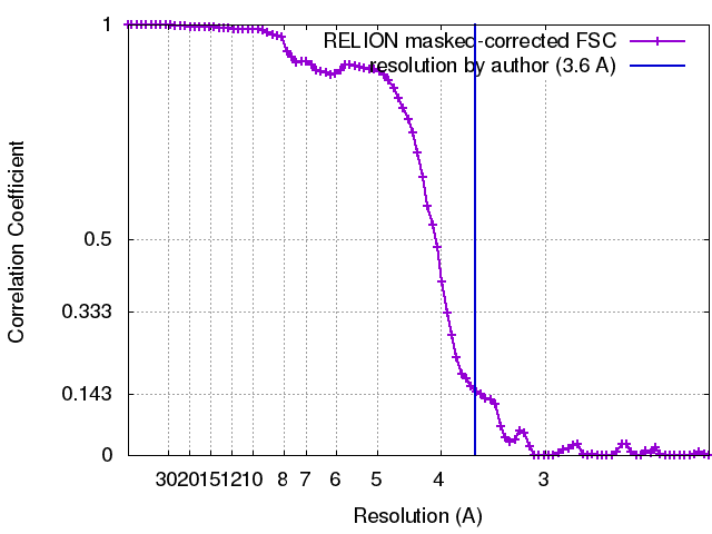
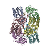
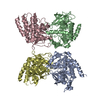
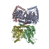
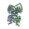
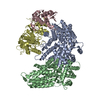
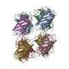
Journal: EMBO J / Year: 2018 Title: Structural basis for assembly of the CBF3 kinetochore complex. Authors: Vera Leber / Andrea Nans / Martin R Singleton / Abstract: Eukaryotic chromosomes contain a specialised region known as the centromere, which forms the platform for kinetochore assembly and microtubule attachment. The centromere is distinguished by the ...Eukaryotic chromosomes contain a specialised region known as the centromere, which forms the platform for kinetochore assembly and microtubule attachment. The centromere is distinguished by the presence of nucleosomes containing the histone H3 variant, CENP-A. In budding yeast, centromere establishment begins with the recognition of a specific DNA sequence by the CBF3 complex. This in turn facilitates CENP-A nucleosome deposition and kinetochore assembly. Here, we describe a 3.6 Å single-particle cryo-EM reconstruction of the core CBF3 complex, incorporating the sequence-specific DNA-binding protein Cep3 together with regulatory subunits Ctf13 and Skp1. This provides the first structural data on Ctf13, defining it as an F-box protein of the leucine-rich-repeat family, and demonstrates how a novel F-box-mediated interaction between Ctf13 and Skp1 is responsible for initial assembly of the CBF3 complex.
History
Deposition
Nov 17, 2017
-
Header (metadata) release
Dec 13, 2017
-
Map release
Dec 13, 2017
-
Update
Dec 11, 2019
-
Current status
Dec 11, 2019
Processing site: PDBe / Status: Released
-
Structure visualization
Movie
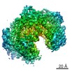
Surface view with section colored by density value
In the structure databanks used in Yorodumi, some data are registered as the other names, "COVID-19 virus" and "2019-nCoV". Here are the details of the virus and the list of structure data.
Jan 31, 2019. EMDB accession codes are about to change! (news from PDBe EMDB page)
EMDB accession codes are about to change! (news from PDBe EMDB page)
The allocation of 4 digits for EMDB accession codes will soon come to an end. Whilst these codes will remain in use, new EMDB accession codes will include an additional digit and will expand incrementally as the available range of codes is exhausted. The current 4-digit format prefixed with “EMD-” (i.e. EMD-XXXX) will advance to a 5-digit format (i.e. EMD-XXXXX), and so on. It is currently estimated that the 4-digit codes will be depleted around Spring 2019, at which point the 5-digit format will come into force.
The EM Navigator/Yorodumi systems omit the EMD- prefix.
Related info.:Q: What is EMD? / ID/Accession-code notation in Yorodumi/EM Navigator
Yorodumi is a browser for structure data from EMDB, PDB, SASBDB, etc.
This page is also the successor to EM Navigator detail page, and also detail information page/front-end page for Omokage search.
The word "yorodu" (or yorozu) is an old Japanese word meaning "ten thousand". "mi" (miru) is to see.
Related info.:EMDB / PDB / SASBDB / Comparison of 3 databanks / Yorodumi Search / Aug 31, 2016. New EM Navigator & Yorodumi / Yorodumi Papers / Jmol/JSmol / Function and homology information / Changes in new EM Navigator and Yorodumi
 Movie
Movie Controller


 Open data
 Basic information
 Map data
 Sample
 Function and homology information
 septin ring assembly / centromeric DNA binding ...RAVE complex / Iron uptake and transport / CBF3 complex / regulation of transcription by galactose / regulation of sulfur amino acid metabolic process / cellular response to methylmercury / vacuolar proton-transporting V-type ATPase complex assembly / FBXL7 down-regulates AURKA during mitotic entry and in early mitosis /
 Authors
 United Kingdom, 3 items
  Citation
 Structure visualization
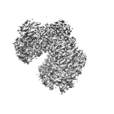
 Downloads & links
 emd_4163.png
 http://ftp.pdbj.org/pub/emdb/structures/EMD-4163


 Sample components
 Processing
 Electron microscopy