 Movie
Movie Controller


+ Open data

- Basic information
| Entry | Database: EMDB / ID: EMD-3953 | |||||||||
|---|---|---|---|---|---|---|---|---|---|---|
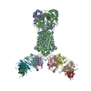
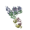
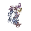
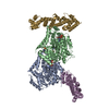
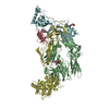
| Title | Structure of inhibitor-bound ABCG2 | |||||||||
 Map data Map data | The local-resolution filtered map of ABCG2-MZ29-Fab | |||||||||
 Sample Sample |
| |||||||||
| Function / homology |  Function and homology information Function and homology informationbiotin transmembrane transporter activity / biotin transport / riboflavin transport / riboflavin transmembrane transporter activity / renal urate salt excretion / urate metabolic process / urate transmembrane transporter activity / Abacavir transmembrane transport / organic anion transport / external side of apical plasma membrane ...biotin transmembrane transporter activity / biotin transport / riboflavin transport / riboflavin transmembrane transporter activity / renal urate salt excretion / urate metabolic process / urate transmembrane transporter activity / Abacavir transmembrane transport / organic anion transport / external side of apical plasma membrane / organic anion transmembrane transporter activity / xenobiotic transport across blood-brain barrier / export across plasma membrane / ABC-type xenobiotic transporter / Paracetamol ADME / Ciprofloxacin ADME / transepithelial transport / cellular detoxification / ABC-type xenobiotic transporter activity / NFE2L2 regulating MDR associated enzymes /  Heme biosynthesis / Heme degradation / lipid transport / xenobiotic transmembrane transporter activity / efflux transmembrane transporter activity / transport across blood-brain barrier / ATPase-coupled transmembrane transporter activity / mitochondrial membrane / brush border membrane / Iron uptake and transport / transmembrane transport / membrane raft / apical plasma membrane / ATP hydrolysis activity / protein homodimerization activity / nucleoplasm / ATP binding / identical protein binding / plasma membrane Heme biosynthesis / Heme degradation / lipid transport / xenobiotic transmembrane transporter activity / efflux transmembrane transporter activity / transport across blood-brain barrier / ATPase-coupled transmembrane transporter activity / mitochondrial membrane / brush border membrane / Iron uptake and transport / transmembrane transport / membrane raft / apical plasma membrane / ATP hydrolysis activity / protein homodimerization activity / nucleoplasm / ATP binding / identical protein binding / plasma membraneSimilarity search - Function | |||||||||
| Biological species |  HOMO SAPIENS (human) / HOMO SAPIENS (human) /  MUS MUSCULUS (house mouse) / Homo sapiens (human) / Mus musculus (house mouse) MUS MUSCULUS (house mouse) / Homo sapiens (human) / Mus musculus (house mouse) | |||||||||
| Method | single particle reconstruction / cryo EM / Resolution: 3.1 Å | |||||||||
 Authors Authors | Jackson SM / Manolaridis I / Kowal J / Zechner M / Altmann KH / Locher KP | |||||||||
| Funding support |  Switzerland, 2 items Switzerland, 2 items
| |||||||||
 Citation Citation | Journal: Nat Struct Mol Biol / Year: 2018 Title: Structural basis of small-molecule inhibition of human multidrug transporter ABCG2. Authors: Scott M Jackson / Ioannis Manolaridis / Julia Kowal / Melanie Zechner / Nicholas M I Taylor / Manuel Bause / Stefanie Bauer / Ruben Bartholomaeus / Guenther Bernhardt / Burkhard Koenig / ...Authors: Scott M Jackson / Ioannis Manolaridis / Julia Kowal / Melanie Zechner / Nicholas M I Taylor / Manuel Bause / Stefanie Bauer / Ruben Bartholomaeus / Guenther Bernhardt / Burkhard Koenig / Armin Buschauer / Henning Stahlberg / Karl-Heinz Altmann / Kaspar P Locher /   Abstract: ABCG2 is an ATP-binding cassette (ABC) transporter that protects tissues against xenobiotics, affects the pharmacokinetics of drugs and contributes to multidrug resistance. Although many inhibitors ...ABCG2 is an ATP-binding cassette (ABC) transporter that protects tissues against xenobiotics, affects the pharmacokinetics of drugs and contributes to multidrug resistance. Although many inhibitors and modulators of ABCG2 have been developed, understanding their structure-activity relationship requires high-resolution structural insight. Here, we present cryo-EM structures of human ABCG2 bound to synthetic derivatives of the fumitremorgin C-related inhibitor Ko143 or the multidrug resistance modulator tariquidar. Both compounds are bound to the central, inward-facing cavity of ABCG2, blocking access for substrates and preventing conformational changes required for ATP hydrolysis. The high resolutions allowed for de novo building of the entire transporter and also revealed tightly bound phospholipids and cholesterol interacting with the lipid-exposed surface of the transmembrane domains (TMDs). Extensive chemical modifications of the Ko143 scaffold combined with in vitro functional analyses revealed the details of ABCG2 interactions with this compound family and provide a basis for the design of novel inhibitors and modulators. | |||||||||
| History |
|
- Structure visualization
| Movie |
Movie viewer |
|---|---|
| Structure viewer | EM map: SurfViewMolmilJmol/JSmol |
| Supplemental images |

- Downloads & links
-EMDB archive
| Map data | emd_3953.map.gz | 132.4 MB | EMDB map data format | |
|---|---|---|---|---|
| Header (meta data) | emd-3953-v30.xmlemd-3953.xml | 17.6 KB 17.6 KB | Display Display | EMDB header |
| FSC (resolution estimation) | emd_3953_fsc.xml | 13.2 KB | Display | FSC data file |
| Images |  emd_3953.png emd_3953.png | 47.4 KB | ||
| Others | emd_3953_additional.map.gz | 14.7 MB | ||
| Archive directory |  http://ftp.pdbj.org/pub/emdb/structures/EMD-3953ftp://ftp.pdbj.org/pub/emdb/structures/EMD-3953 http://ftp.pdbj.org/pub/emdb/structures/EMD-3953ftp://ftp.pdbj.org/pub/emdb/structures/EMD-3953 | HTTPS FTP |
-Related structure data
| Related structure data |  6etiMC 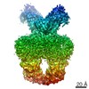 4246C 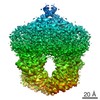 4256C  6feqC  6ffcC  6hijC M: atomic model generated by this map C: citing same article ( |
|---|---|
| Similar structure data | |
| EM raw data | EMPIAR-10374 (Title: CryoEM reconstruction of human ABCG2 transporter with inhibitor MZ29 and 5D3-Fab Data size: 5.6 TB Data #1: Unaligned super-resolution multi-frame micrographs of MZ-29 inhibitor bound ABCG2-Fab. [micrographs - multiframe] Data #2: Extracted particles and corresponding star file [picked particles - single frame - processed]) |


-Links
| EMDB pages | EMDB (EBI/PDBe) / EMDataResource |
|---|---|
| Related items in Molecule of the Month |


-Map
| File | Download / File: emd_3953.map.gz / Format: CCP4 / Size: 216 MB / Type: IMAGE STORED AS FLOATING POINT NUMBER (4 BYTES) | ||||||||||||||||||||||||||||||||||||||||||||||||||||||||||||
|---|---|---|---|---|---|---|---|---|---|---|---|---|---|---|---|---|---|---|---|---|---|---|---|---|---|---|---|---|---|---|---|---|---|---|---|---|---|---|---|---|---|---|---|---|---|---|---|---|---|---|---|---|---|---|---|---|---|---|---|---|---|
| Annotation | The local-resolution filtered map of ABCG2-MZ29-Fab | ||||||||||||||||||||||||||||||||||||||||||||||||||||||||||||
| Voxel size | X=Y=Z: 0.84 Å | ||||||||||||||||||||||||||||||||||||||||||||||||||||||||||||
| Density |
| ||||||||||||||||||||||||||||||||||||||||||||||||||||||||||||
| Symmetry | Space group: 1 | ||||||||||||||||||||||||||||||||||||||||||||||||||||||||||||
| Details | EMDB XML:
CCP4 map header:
| ||||||||||||||||||||||||||||||||||||||||||||||||||||||||||||
-Supplemental data
-Additional map: The post-processed map of ABCG2-MZ29-Fab
| File | emd_3953_additional.map | ||||||||||||
|---|---|---|---|---|---|---|---|---|---|---|---|---|---|
| Annotation | The post-processed map of ABCG2-MZ29-Fab | ||||||||||||
| Projections & Slices |
| ||||||||||||
| Density Histograms |
 Z
Z Y
Y X
X


- Sample components
-Entire : inhibitor-bound ABCG2
| Entire | Name: inhibitor-bound ABCG2 |
|---|---|
| Components |
|
-Supramolecule #1: inhibitor-bound ABCG2
| Supramolecule | Name: inhibitor-bound ABCG2 / type: complex / ID: 1 / Parent: 0 / Macromolecule list: #2-#3 |
|---|
-Supramolecule #2: ABCG2
| Supramolecule | Name: ABCG2 / type: complex / ID: 2 / Parent: 1 / Macromolecule list: #1 |
|---|---|
| Source (natural) | Organism: HOMO SAPIENS (human) |
-Supramolecule #3: inhibitor
| Supramolecule | Name: inhibitor / type: complex / ID: 3 / Parent: 1 / Macromolecule list: #2-#3 |
|---|---|
| Source (natural) | Organism: MUS MUSCULUS (house mouse) |
-Macromolecule #1: ATP-binding cassette sub-family G member 2
| Macromolecule | Name: ATP-binding cassette sub-family G member 2 / type: protein_or_peptide / ID: 1 / Number of copies: 2 / Enantiomer: LEVO |
|---|---|
| Source (natural) | Organism: Homo sapiens (human) |
| Molecular weight | Theoretical: 72.385852 KDa |
| Recombinant expression | Organism: Homo sapiens (human) |
| Sequence | String: MSSSNVEVFI PVSQGNTNGF PATASNDLKA FTEGAVLSFH NICYRVKLKS GFLPCRKPVE KEILSNINGI MKPGLNAILG PTGGGKSSL LDVLAARKDP SGLSGDVLIN GAPRPANFKC NSGYVVQDDV VMGTLTVREN LQFSAALRLA TTMTNHEKNE R INRVIQEL ...String: MSSSNVEVFI PVSQGNTNGF PATASNDLKA FTEGAVLSFH NICYRVKLKS GFLPCRKPVE KEILSNINGI MKPGLNAILG PTGGGKSSL LDVLAARKDP SGLSGDVLIN GAPRPANFKC NSGYVVQDDV VMGTLTVREN LQFSAALRLA TTMTNHEKNE R INRVIQEL GLDKVADSKV GTQFIRGVSG GERKRTSIGM ELITDPSILF LDEPTTGLDS STANAVLLLL KRMSKQGRTI IF SIHQPRY SIFKLFDSLT LLASGRLMFH GPAQEALGYF ESAGYHCEAY NNPADFFLDI INGDSTAVAL NREEDFKATE IIE PSKQDK PLIEKLAEIY VNSSFYKETK AELHQLSGGE KKKKITVFKE ISYTTSFCHQ LRWVSKRSFK NLLGNPQASI AQII VTVVL GLVIGAIYFG LKNDSTGIQN RAGVLFFLTT NQCFSSVSAV ELFVVEKKLF IHEYISGYYR VSSYFLGKLL SDLLP MRML PSIIFTCIVY FMLGLKPKAD AFFVMMFTLM MVAYSASSMA LAIAAGQSVV SVATLLMTIC FVFMMIFSGL LVNLTT IAS WLSWLQYFSI PRYGFTALQH NEFLGQNFCP GLNATGNNPC NYATCTGEEY LVKQGIDLSP WGLWKNHVAL ACMIVIF LT IAYLKLLFLK KYS |
-Macromolecule #2: 5D3(Fab) light chain variable domain
| Macromolecule | Name: 5D3(Fab) light chain variable domain / type: protein_or_peptide / ID: 2 / Details: The variable domain of the light chain of 5D3(Fab) / Number of copies: 2 / Enantiomer: LEVO |
|---|---|
| Source (natural) | Organism: Mus musculus (house mouse) |
| Molecular weight | Theoretical: 23.594016 KDa |
| Recombinant expression | Organism: Mus musculus (house mouse) |
| Sequence | String: DIVLTQSPSS FSVSLGDRVT ISCKASGYIL NRLAWYQQKP GNAPRLLISG ATSLETGFPS RFSGTGSGKD YTLSISSLQT EDVGTYYCQ QYWSTPWTFG GGTKLEIRRA DAAPTVSIFP PSSEQLTSGG ASVVCFLNNF YPKDINVKWK IDGSERQNGV L NSWTDQDS ...String: DIVLTQSPSS FSVSLGDRVT ISCKASGYIL NRLAWYQQKP GNAPRLLISG ATSLETGFPS RFSGTGSGKD YTLSISSLQT EDVGTYYCQ QYWSTPWTFG GGTKLEIRRA DAAPTVSIFP PSSEQLTSGG ASVVCFLNNF YPKDINVKWK IDGSERQNGV L NSWTDQDS KDSTYSMSST LTLTKDEYER HNSYTCEATH KTSTSPIVKS FNRNEC |
-Macromolecule #3: 5D3(Fab) heavy chain variable domain
| Macromolecule | Name: 5D3(Fab) heavy chain variable domain / type: protein_or_peptide / ID: 3 / Details: The variable domain of the heavy chain of 5D3(Fab) / Number of copies: 2 / Enantiomer: LEVO |
|---|---|
| Source (natural) | Organism: Mus musculus (house mouse) |
| Molecular weight | Theoretical: 23.843633 KDa |
| Recombinant expression | Organism: Mus musculus (house mouse) |
| Sequence | String: QVQLQESGPG LVKPSQSLSL TCTVTGFSIT SDYAWNWIRQ FPGKKLEWMG YINFDGGTTY NPSLRGRISI TRDTSKNQFF LQLRSVTPE DTATYYCATF YGAKGTLDYW GQGTSVTVSS AKTTPPSVYP LAPVCGDTSG SSVTLGCLVK GYFPEPVTLT W NSGSLSSG ...String: QVQLQESGPG LVKPSQSLSL TCTVTGFSIT SDYAWNWIRQ FPGKKLEWMG YINFDGGTTY NPSLRGRISI TRDTSKNQFF LQLRSVTPE DTATYYCATF YGAKGTLDYW GQGTSVTVSS AKTTPPSVYP LAPVCGDTSG SSVTLGCLVK GYFPEPVTLT W NSGSLSSG VHTFPAVLQS DLYTLSSSVT VTSSTWPSQS ITCNVAHPAS STKVDKKIEP RGP |
-Macromolecule #5: ~{tert}-butyl 3-[(2~{S},5~{S},8~{S})-14-cyclopentyloxy-2-(2-methy...
| Macromolecule | Name: ~{tert}-butyl 3-[(2~{S},5~{S},8~{S})-14-cyclopentyloxy-2-(2-methylpropyl)-4,7-bis(oxidanylidene)-3,6,17-triazatetracyclo[8.7.0.0^{3,8}.0^{11,16}]heptadeca-1(10),11,13,15-tetraen-5-yl]propanoate type: ligand / ID: 5 / Number of copies: 2 / Formula: BWQ |
|---|---|
| Molecular weight | Theoretical: 523.664 Da |
| Chemical component information |  ChemComp-BWQ: |
-Experimental details
-Structure determination
| Method | cryo EM |
|---|---|
 Processing Processing | single particle reconstruction |
| Aggregation state | particle |
-Sample preparation
| Concentration | 0.4 mg/mL |
|---|---|
| Buffer | pH: 7.5 |
| Grid | Model: Quantifoil R1.2/1.3 / Material: COPPER / Mesh: 300 / Pretreatment - Type: GLOW DISCHARGE |
| Vitrification | Cryogen name: ETHANE-PROPANE / Chamber humidity: 100 % / Chamber temperature: 277 K / Instrument: FEI VITROBOT MARK IV |
- Electron microscopy
| Microscope | FEI TITAN KRIOS |
|---|---|
| Electron beam | Acceleration voltage: 300 kV / Electron source: FIELD EMISSION GUN |
| Electron optics | Illumination mode: FLOOD BEAM / Imaging mode: BRIGHT FIELDBright-field microscopy |
| Image recording | Film or detector model: GATAN K2 SUMMIT (4k x 4k) / Number real images: 4094 / Average exposure time: 0.2 sec. / Average electron dose: 2.0 e/Å2 |
| Experimental equipment |  Model: Titan Krios / Image courtesy: FEI Company |
-Image processing
| Startup model | Type of model: PDB ENTRY / Details: 5NJ3 |
|---|---|
| Initial angle assignment | Type: PROJECTION MATCHING |
| Final angle assignment | Type: PROJECTION MATCHING |
| Final reconstruction | Resolution.type: BY AUTHOR / Resolution: 3.1 Å / Resolution method: FSC 0.143 CUT-OFF / Software - Name: RELION / Number images used: 284831 |
| FSC plot (resolution estimation) |  |
-Atomic model buiding 1
| Refinement | Overall B value: 98 |
|---|---|
| Output model | PDB-6eti: |
