 Movie
Movie Controller

[English] 日本語
 Yorodumi
- EMDB-3869: The structure of Ebola virus nucleoprotein (residues 1-450) in nu... -
+ Open data

- Basic information
| Entry | Database: EMDB / ID: EMD-3869 | ||||||||||||
|---|---|---|---|---|---|---|---|---|---|---|---|---|---|
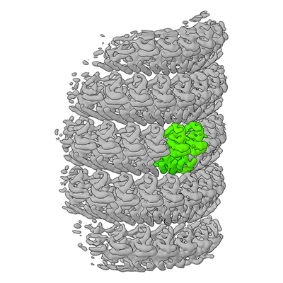
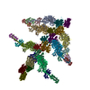
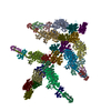
| Title | The structure of Ebola virus nucleoprotein (residues 1-450) in nucleocapsid-like assemblies | ||||||||||||
 Map data Map data | Subtomogram averaging of recombinantly expressed Ebola Nucleoprotein (NP), residues 1-450 | ||||||||||||
 Sample Sample |
| ||||||||||||
| Function / homology | Ebola nucleoprotein / Ebola nucleoprotein / viral RNA genome packaging / helical viral capsid / viral nucleocapsid / host cell cytoplasm /  ribonucleoprotein complex / RNA binding / Nucleoprotein ribonucleoprotein complex / RNA binding / Nucleoprotein Function and homology information Function and homology information | ||||||||||||
| Biological species |   Zaire ebolavirus (strain Mayinga-76) / Ebola virus - Mayinga, Zaire, 1976 Zaire ebolavirus (strain Mayinga-76) / Ebola virus - Mayinga, Zaire, 1976 | ||||||||||||
| Method | subtomogram averaging / cryo EM / Resolution: 6.6 Å | ||||||||||||
 Authors Authors | Wan W / Kolesnikova L / Clarke M / Koehler A / Noda T / Becker S / Briggs JAG | ||||||||||||
| Funding support |  Germany, 3 items Germany, 3 items
| ||||||||||||
 Citation Citation | Journal: Nature / Year: 2017 Title: Structure and assembly of the Ebola virus nucleocapsid. Authors: William Wan / Larissa Kolesnikova / Mairi Clarke / Alexander Koehler / Takeshi Noda / Stephan Becker / John A G Briggs /   Abstract: Ebola and Marburg viruses are filoviruses: filamentous, enveloped viruses that cause haemorrhagic fever. Filoviruses are within the order Mononegavirales, which also includes rabies virus, measles ...Ebola and Marburg viruses are filoviruses: filamentous, enveloped viruses that cause haemorrhagic fever. Filoviruses are within the order Mononegavirales, which also includes rabies virus, measles virus, and respiratory syncytial virus. Mononegaviruses have non-segmented, single-stranded negative-sense RNA genomes that are encapsidated by nucleoprotein and other viral proteins to form a helical nucleocapsid. The nucleocapsid acts as a scaffold for virus assembly and as a template for genome transcription and replication. Insights into nucleoprotein-nucleoprotein interactions have been derived from structural studies of oligomerized, RNA-encapsidating nucleoprotein, and cryo-electron microscopy of nucleocapsid or nucleocapsid-like structures. There have been no high-resolution reconstructions of complete mononegavirus nucleocapsids. Here we apply cryo-electron tomography and subtomogram averaging to determine the structure of Ebola virus nucleocapsid within intact viruses and recombinant nucleocapsid-like assemblies. These structures reveal the identity and arrangement of the nucleocapsid components, and suggest that the formation of an extended α-helix from the disordered carboxy-terminal region of nucleoprotein-core links nucleoprotein oligomerization, nucleocapsid condensation, RNA encapsidation, and accessory protein recruitment. | ||||||||||||
| History |
|
- Structure visualization
| Movie |
Movie viewer |
|---|---|
| Structure viewer | EM map: SurfViewMolmilJmol/JSmol |
| Supplemental images |
- Downloads & links
-EMDB archive
| Map data | emd_3869.map.gz | 23.9 MB | EMDB map data format | |
|---|---|---|---|---|
| Header (meta data) | emd-3869-v30.xmlemd-3869.xml | 17.8 KB 17.8 KB | Display Display | EMDB header |
| Images |  emd_3869.png emd_3869.png | 140.8 KB | ||
| Archive directory |  http://ftp.pdbj.org/pub/emdb/structures/EMD-3869ftp://ftp.pdbj.org/pub/emdb/structures/EMD-3869 http://ftp.pdbj.org/pub/emdb/structures/EMD-3869ftp://ftp.pdbj.org/pub/emdb/structures/EMD-3869 | HTTPS FTP |
-Related structure data
| Related structure data | 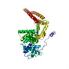 6ehlMC  3870C 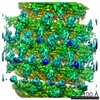 3871C 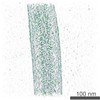 3872C 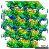 3873C 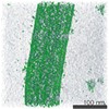 3874C 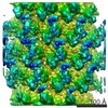 3875C 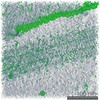 3876C 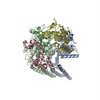 6ehmC M: atomic model generated by this map C: citing same article ( |
|---|---|
| Similar structure data |


-Links
| EMDB pages | EMDB (EBI/PDBe) / EMDataResource |
|---|---|
| Related items in Molecule of the Month |

-Map
| File | Download / File: emd_3869.map.gz / Format: CCP4 / Size: 27 MB / Type: IMAGE STORED AS FLOATING POINT NUMBER (4 BYTES) | ||||||||||||||||||||||||||||||||||||||||||||||||||||||||||||
|---|---|---|---|---|---|---|---|---|---|---|---|---|---|---|---|---|---|---|---|---|---|---|---|---|---|---|---|---|---|---|---|---|---|---|---|---|---|---|---|---|---|---|---|---|---|---|---|---|---|---|---|---|---|---|---|---|---|---|---|---|---|
| Annotation | Subtomogram averaging of recombinantly expressed Ebola Nucleoprotein (NP), residues 1-450 | ||||||||||||||||||||||||||||||||||||||||||||||||||||||||||||
| Voxel size | X=Y=Z: 1.78 Å | ||||||||||||||||||||||||||||||||||||||||||||||||||||||||||||
| Density |
| ||||||||||||||||||||||||||||||||||||||||||||||||||||||||||||
| Symmetry | Space group: 1 | ||||||||||||||||||||||||||||||||||||||||||||||||||||||||||||
| Details | EMDB XML:
CCP4 map header:
| ||||||||||||||||||||||||||||||||||||||||||||||||||||||||||||
-Supplemental data
- Sample components
-Entire : Ebola virus - Mayinga, Zaire, 1976
| Entire | Name: Ebola virus - Mayinga, Zaire, 1976 |
|---|---|
| Components |
|
-Supramolecule #1: Ebola virus - Mayinga, Zaire, 1976
| Supramolecule | Name: Ebola virus - Mayinga, Zaire, 1976 / type: virus / ID: 1 / Parent: 0 / Macromolecule list: all / NCBI-ID: 128952 / Sci species name: Ebola virus - Mayinga, Zaire, 1976 / Virus type: VIRUS-LIKE PARTICLE / Virus isolate: STRAIN / Virus enveloped: No / Virus empty: No |
|---|---|
| Host system | Organism:  Homo sapiens (human) / Recombinant cell: HEK 293T Homo sapiens (human) / Recombinant cell: HEK 293T |
| Virus shell | Shell ID: 1 / Name: Nucleocapsid / Diameter: 280.0 Å |
-Macromolecule #1: Nucleoprotein
| Macromolecule | Name: Nucleoprotein / type: protein_or_peptide / ID: 1 / Number of copies: 1 / Enantiomer: LEVO |
|---|---|
| Source (natural) | Organism: Zaire ebolavirus (strain Mayinga-76) / Strain: Mayinga-76 |
| Molecular weight | Theoretical: 83.3875 KDa |
| Recombinant expression | Organism: Homo sapiens (human) |
| Sequence | String: MDSRPQKIWM APSLTESDMD YHKILTAGLS VQQGIVRQRV IPVYQVNNLE EICQLIIQAF EAGVDFQESA DSFLLMLCLH HAYQGDYKL FLESGAVKYL EGHGFRFEVK KRDGVKRLEE LLPAVSSGKN IKRTLAAMPE EETTEANAGQ FLSFASLFLP K LVVGEKAC ...String: MDSRPQKIWM APSLTESDMD YHKILTAGLS VQQGIVRQRV IPVYQVNNLE EICQLIIQAF EAGVDFQESA DSFLLMLCLH HAYQGDYKL FLESGAVKYL EGHGFRFEVK KRDGVKRLEE LLPAVSSGKN IKRTLAAMPE EETTEANAGQ FLSFASLFLP K LVVGEKAC LEKVQRQIQV HAEQGLIQYP TAWQSVGHMM VIFRLMRTNF LIKFLLIHQG MHMVAGHDAN DAVISNSVAQ AR FSGLLIV KTVLDHILQK TERGVRLHPL ARTAKVKNEV NSFKAALSSL AKHGEYAPFA RLLNLSGVNN LEHGLFPQLS AIA LGVATA HGSTLAGVNV GEQYQQLREA ATEAEKQLQQ YAESRELDHL GLDDQEKKIL MNFHQKKNEI SFQQTNAMVT LRKE RLAKL TEAITAASLP KTSGHYDDDD DIPFPGPIND DDNPGHQDDD PTDSQDTTIP DVVVDPDDGS YGEYQSYSEN GMNAP DDLV LFDLDEDDED TKPVPNRSTK GGQQKNSQKG QHIEGRQTQS RPIQNVPGPH RTIHHASAPL TDNDRRNEPS GSTSPR MLT PINEEADPLD DADDETSSLP PLESDDEEQD RDGTSNRTPT VAPPAPVYRD HSEKKELPQD EQQDQDHTQE ARNQDSD NT QSEHSFEEMY RHILRSQGPF DAVLYYHMMK DEPVVFSTSD GKEYTYPDSL EEEYPPWLTE KEAMNEENRF VTLDGQQF Y WPVMNHKNKF MAILQHHQ |
-Experimental details
-Structure determination
| Method | cryo EM |
|---|---|
 Processing Processing | subtomogram averaging |
| Aggregation state | helical array |
-Sample preparation
| Buffer | pH: 7.4 Component:
| |||||||||||||||
|---|---|---|---|---|---|---|---|---|---|---|---|---|---|---|---|---|
| Grid | Model: C-flat 2/1 3C / Material: COPPER / Mesh: 300 / Support film - Material: CARBON / Support film - topology: HOLEY / Support film - Film thickness: 20.0 nm / Pretreatment - Type: GLOW DISCHARGE / Pretreatment - Atmosphere: AIR / Pretreatment - Pressure: 0.039 kPa | |||||||||||||||
| Vitrification | Cryogen name: ETHANE / Chamber humidity: 95 % / Instrument: FEI VITROBOT MARK II |
- Electron microscopy
| Microscope | FEI TITAN KRIOS |
|---|---|
| Electron beam | Acceleration voltage: 300 kV / Electron source: FIELD EMISSION GUN |
| Electron optics | C2 aperture diameter: 50.0 µm / Illumination mode: FLOOD BEAM / Imaging mode: BRIGHT FIELDBright-field microscopy / Cs: 2.7 mm / Nominal defocus max: 4.5 µm / Nominal defocus min: 2.0 µm / Nominal magnification: 81000 |
| Specialist optics | Energy filter - Name: GIF Quantum LS / Energy filter - Lower energy threshold: -10 eV / Energy filter - Upper energy threshold: 10 eV |
| Sample stage | Specimen holder model: FEI TITAN KRIOS AUTOGRID HOLDER / Cooling holder cryogen: NITROGEN |
| Image recording | Film or detector model: GATAN K2 QUANTUM (4k x 4k) / Detector mode: SUPER-RESOLUTION / Digitization - Dimensions - Width: 3708 pixel / Digitization - Dimensions - Height: 3708 pixel / Digitization - Frames/image: 1-5 / Number grids imaged: 1 / Average exposure time: 1.0 sec. / Average electron dose: 2.4 e/Å2 |
| Experimental equipment |  Model: Titan Krios / Image courtesy: FEI Company |
-Image processing
| Extraction | Number tomograms: 63 / Number images used: 488916 / Reference model: None Software:
Details: Points along the helical axis were manually placed to define a spline. A cylindrical grid as defined at a given radius from the spline; grid spacing was chosen to provide ~4x oversampling. | ||||||
|---|---|---|---|---|---|---|---|
| CTF correction | Software:
Details: CTF amplitude correction was performed during the wedge-weighted subtomogram averaging step. | ||||||
| Final angle assignment | Type: OTHER / Software - Name: AV3 Details: Iterative angular search was performed via maximization of a modified constrained cross-correlation function. | ||||||
| Final reconstruction | Number classes used: 1 / Applied symmetry - Point group: C1 (asymmetric) / Algorithm: BACK PROJECTION / Resolution.type: BY AUTHOR / Resolution: 6.6 Å / Resolution method: FSC 0.143 CUT-OFF / Software - Name: AV3 Details: Local resolution was estimated using moving window FSC calculations. Number subtomograms used: 1 | ||||||
| Details | Frames were aligned using K2Align software, based off the MotionCorr algorithm. Tomograms were reconstructed with IMOD, using stripwise CTF-correction and weighted back projection. Subtomogram averaging was performed using scripts derived from TOM, AV3, and DYNAMO. |
-Atomic model buiding 1
| Details | Crystal structure was rigid-body fitted into density using Chimera. Missing regions of the structure was built using ideal secondary structures in coot. These were then flexibly fit using MDFF and NAMD. |
|---|---|
| Refinement | Protocol: FLEXIBLE FIT |
| Output model | PDB-6ehl: |
